Journal of
eISSN: 2373-6410


Case Report Volume 5 Issue 3
1Department of Neurological Surgery, Hospital de Alta Complejidad Juan D Perón, Argentina
2Department of Neurological Surgery, Hospital Juan A Fernández, Argentina
3Department of Anaesthesia, Hospital de Alta Complejidad Juan D Perón, Argentina
Correspondence: Mannará Francisco Alberto, Department of Neurological Surgery. Hospital Juan A Fernández, Avalos 1536, (1431), Buenos Aires, Argentina, Tel 5491154996753
Received: October 22, 2016 | Published: November 11, 2016
Citation: Mannara FA, Alonso S, Gonza H, Fernández J, Stupka S, et al. (2016) Use of Adenosine in Cerebral Aneurysm Surgeries. J Neurol Stroke 5(3): 00177. DOI: DOI: 10.15406/jnsk.2016.05.00177
Transient flow arrest caused by induced Adenonsine administration has been used to facilitate microsurgical clipping in some cases of cerebral aneurysms. Here, we described our experience in two cases where adenosine induced flow arrest facilitate to place the clip in the neck of aneurysm without any complication, in cases where proximal control is not possible.
Keywords: Adenosine, Flow arrest, Cerebral aneurysm, Aneurysm clipping
Endovascular techniques in the treatment of cerebral aneurysms had evolved advances in the field, however, microsurgery with clip occlusion of the aneurysm´s neck is still a mainstay in definitive treatment. The rupture of cerebral aneurysm during the surgery may cause death or permanent disability. To avoid rupture, transient clipping proximal to the aneurysm is usually used to decrease the turgor of the aneurysm neck. Sometimes there are cases, as carotid paraclinoid aneurysm or giant aneurysm even with clinoidectomy performed, where it is hard to find an anatomically right place to make the temporary arterial occlusion. In such cases, the use of adenosine can facilitate surgery, producing reversible flow arrest; so that the volume of aneurysm sac decreases letting a better visual image to set down the clip. We present two cases where the use of adenosine-induced flow arrest facilitates placement of the clip in the prompt position without any complication.
Case 1
51 years old female patient, antecedent of endovascular coiled left paraclinoid aneurysm 2 years before, with subsequent amaurosis. She was admitted to hospital with a ruptured carotid ophthalmic on the right side (Figure 1). She was listed for surgical clipping of the aneurysm. To prevent intra-operative rupture we planned to use intravenous adenosine. Whenever we use this type of technique, we prepare the patient with external defibrillator paddles in the chest.
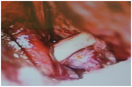
We performed a right frontotemporal craniotomy with intradural clinoidectomy. After the aneurysm neck was exposed, a great tension on the artery wall was confirmed, with the optic nerve displaced upwards, so it was difficult to clip the aneurysm safely (Figure 2). Adenosine 18 mg (0.03 mg/kg) was given via central venous catheter in a quick bolus, producing flow arrest and transient a systole for 20 seconds. This maneuver produced the softening of the dome and neck (Figure 3) and facilitated the clipping. Exclusion of aneurysm was confirmed with Doppler ultrasonographic device (20 MHZ Mizuho Doppler). The patient did not experienced any miocardial or neurological issues. ECG, echocardiogram and troponin I remained within normal limits 6 and 24 hours after surgery (Figure 4).

Figure 2 Intraoperative. Before adenosine-induced flow arrest. Aneurysm with increased tension in the wall.
Case 2
35 years old patient with subarachnoid haemorrhage, Hunt and Hess grade IV, Fisher 4, with left giant supraclinoid carotid aneurysm (Figure 5). Clip reconstruction and use of adenosine was planned in this case. During surgical dissection, the dome was ruptured producing cataclysmic bleeding.
Adenosine was infused producing flow arrest, allowing us to perform the reconstruction with fenestrated multiclipping technique in the parent artery, with exclusion of aneurysm. Patency of the internal carotid artery was confirmed with Mizuho Doppler.
Adenosine is an endogenous purine, that slows down the heart rate by acting through the sinoatrial and atrioventricular nodes. Its half life is less than 10 seconds.1 The administration of adenosine in patients with normal sinus rhythm induces a rapidly reversible cardiac arrest. The intravascular half-life of adenosine at physiological level is less than a second.1-3
Adenosine-induced flow arrest is a procedure that diminishes cerebral perfusion pressure during a short and controlled time facilitating the clipping.4,5 In case number 1 we used adenosine because of the great tension on the wall of the aneurysm. We had previously opened the aneurysm in former surgeries and deflated it to perform the clipping; but this option was used when the wall was calcified or affected by thrombosis. The current patient was young and didn´t have a calcified wall, so adenosine-induced flow arrest facilitated the clipping, by softing the walls without the need to retract the optic nerve.
If an aneurysm rupture occurs during surgery, adenosine has been used to produce transient cardiac arrest to stop the bleeding, clarifying the surgical field in order to optimize the clipping.6-10
In our experience, when dealing with an intraoperative aneurysm rupture, a transient clip is used to gain proximal control. Furthermore, lowering systolic pressure facilitates handling this situation. In such circumstances, adenosine may not be used.
In case number 2, we didn´t have proximal control to manage the bleeding. In such circumstance, adenosine-induced flow arrest facilitated the aneurysm clipping. However, the surgeon should think in this possibility to prepare the patient prior to perform surgery, (e.g. external defibrillator pads). Furthermore monitoring all these patients for biochemical evidence of myocardial injury with troponin I in the postoperative period is mandatory to treat any possible complication and improve the result of the procedure.
The use of adenosine in the surgical treatment of cerebral aneurysms in which temporary occlusion is hard or impossible gives a safe choice to simplify microsurgical clipping.
None.
None.

©2016 Mannara, et al. This is an open access article distributed under the terms of the, which permits unrestricted use, distribution, and build upon your work non-commercially.