Journal of
eISSN: 2373-6410


Clinical Images Volume 10 Issue 2
Degenerative and Vascular Cognitive Disorders, Université de Lille 2, France
Correspondence: Jacques De Reuck, Degenerative and Vascular Cognitive Disorders, Université de Lille 2, INSERM U1171, Lille, France
Received: December 10, 2019 | Published: April 24, 2020
Citation: Reuck JD. Post-mortem 7.0-tesla magnetic resonance imaging of polymicrogyria. J Neurol Stroke. 2020;10(2):78. DOI: 10.15406/jnsk.2020.10.00415
Polymicrogyria (PMG) is a malformation of cortical folding with disruption of the pialimitans associated with glial and macrophage accumulation.1 On magnetic resonance imaging of patients with PMG two patterns are observed: one with small fine, and undulating cortex with normal thickness and another with bumpy and abnormally thick cortex.2 Also different types of hippocampus malformations can be associated to PMG.3 7-tesla MRI has main advances compared to those with 1.5 and 3-tesla.4
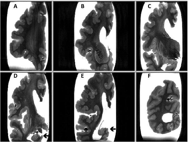
We present a post-mortem 7.0-Tesla magnetic resonance imaging (MRI) study of a 70-year old man with a history of epilepsy, due to PMG and suspected hippocampal sclerosis. Six coronal sections of a cerebral hemisphere and a horizontal section of a cerebellar hemisphere were examined with T2 and T2* sequences. PMG was mainly observed in the parietal cortex. The hippocampus had a normal aspect. Moderate white matter changes (WMC) were present mainly in the parietal region. No cortical micro-infarcts were observed (Figure 1). On the T2* sequence only a few cortical micro-bleeds (CoMBs) were present. The cerebellar cortex had a normal appearance.

Figure 1 On the T2 MRI sequence polymicrogyria is mainly observed in the parietical cortex (sections D and F). The hippocampus has a normal appearance (arrows).
This is the first post-mortem study 7.0-tesla MRI study of PMG that confirms the validity of the “in vivo” imaging. The present case shows mainly parietal gyral malformations. The WMC and CoMBs are to be considered as age-related changes, as observed in normal elderly brains.5
None.
The author declares no conflict of interest.

©2020 Reuck. This is an open access article distributed under the terms of the, which permits unrestricted use, distribution, and build upon your work non-commercially.