Journal of
eISSN: 2373-6410


Short Communication Volume 14 Issue 4
1Student B.Tech (BT), NIIT University, Rajasthan, India
2Department of Neurology, Pushpawati Singhania Research Institute, India
Correspondence: Nitin K Sethi, MD, MBBS, FAAN, PSRI, Hospital, New Delhi, India
Received: July 10, 2024 | Published: July 24, 2024
Citation: Batra D, Sethi NK. Non physiological artifacts in EEG laboratory of a tertiary level hospital in New Delhi. J Neurol Stroke. 2024;14(4):88-90. DOI: 10.15406/jnsk.2024.14.00589
Electroencephalogram (EEG) is a safe and widely used diagnostic test that records the brain's spontaneous electrical activity. It helps detect potential brain anomalies with the highest utility in identification and characterization of seizure disorder. Artifacts frequently contaminate EEG record obscuring the underlying waveforms. Artifacts are signals not originating from the brain and are broadly classified as either physiological or non-physiological. Physiological artifacts arise from the patient and include cardiac, pulse, respiratory, eye movement, and muscle movement artifacts among others. Non-physiological artifacts commonly arise from the patient’s surroundings. Electric line interference, electrode pop, cable movement and bad channel connection can all contaminate the record. We investigated non-physiological artifacts in the outpatient EEG laboratory of a tertiary care hospital in New Delhi and propose suggestions to reduce these artifacts to allow accurate interpretation of EEG record.
Keywords: electroencephalography, non-physiological artifacts, mains interference, electrode pop, eeg laboratory
Surface EEG is a noninvasive neurophysiological technique with the greatest utility in identification and characterization of seizure disorder. It is also a useful test to assess for and grade the degree of cerebral dysfunction (encephalopathy). EEG has the advantage of non-invasiveness, with no exposure to radiation. This portability, affordability, and ability to capture rapid electrical changes make EEG the preferred choice for diagnosing many neurological conditions. EEG measures the combined electrical activity of large groups of neurons, typically in the microvolt range. This information proves valuable in various fields like neuroscience, psychology, cognitive science, and psychophysiology in clinical and research settings. Among clinical neurological conditions, EEG finds utility in sleep disorders, depression, epilepsy, dementia, functional neurological disorders, movement disorders, and schizophrenia among others. While EEG offers numerous benefits, it's not without limitations. A major hurdle is the presence of artifacts. These can either originate from the patient itself (referred to as physiological artifacts) or from patient’s immediate surroundings (referred to as non-physiological artifacts). These artifacts contaminate the EEG making analysis and interpretation of waveforms challenging. A novice EEG reader may misinterpret these artifacts as potential epileptiform discharges leading to inaccurate diagnostic conclusions. Therefore it is imperative that an effort is made to identify and eliminate these artifacts.
We investigated the potential source of non-physiological artifacts in the outpatient EEG laboratory of our hospital. PSRI hospital is a 200 bed tertiary care hospital in New Delhi. The 40-50 square meter (430-538 sq ft) Neurosciences laboratory is functionally divided into two sections. One section houses dedicated setups for EMG measurements, while the other accommodates two EEG machines, one from Medicaid (Medicaid Systems, India) and the other unit is from Neurosoft (Neurosoft, Ivanovo, Russia) (Figure 1).
We investigated the potential sources of non-physiological artifacts in EEG recordings. Upon visual inspection of EEG studies, various non-physiological artifacts were identified. To understand their potential source, a comprehensive investigation was conducted of the outpatient EEG laboratory. Several potential sources of non-physiological artifacts were identified.

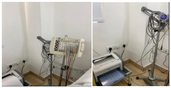
Figure 2 The image portrays potential source of power fluctuations in a hospital room. The EEG machine is positioned next to a printer. Both devices are plugged into the same power outlet. This setup can introduce electrical noise into the EEG signal. In clinical settings, dedicated outlets or surge protectors are recommended to minimize interference and ensure reliable EEG recordings.
Our study goal was to improve the quality and reliability of EEG recordings in the outpatient EEG laboratory of our hospital by identifying the source of non-physiological artifacts and their elimination. We identified several sources of non-physiological artifacts as detailed above.
Power fluctuations can be mitigated by implementing proper grounding techniques and voltage regulators.1,2 Additionally, securely fixing the photic stimulator during VEP studies minimizes movement artifacts.3,4 Utilization of Faraday cages or shielded rooms specifically designed for EEG recordings significantly reduces external electrical interference from sources like power lines and fluorescent lights.1,2,5 Strict policy should be enforced disallowing use of electronic devices like personal cellphones within the EEG recording area.1,2
Modern EEG amplifiers equipped with high input impedance effectively address high electrode impedance, allowing for better capture of weak brain signals through the scalp's resistance.1,2 Regular maintenance and calibration of the EEG machine, including increasing the calibration frequency to at least four times a week, further minimizes the risk of artifacts contaminating the recordings.1,2 High quality, well-maintained filters with sharp cut-off points alongside regular maintenance optimizes filter performance and minimizes signal attenuation during noise removal.1,2,6
Each of the above non-physiological artifacts has the potential to render parts of the record or the entire record difficult to interpret. One should also not forget that a given EEG record may be contaminated by multiple non-physiological artifacts. Our study highlights the importance of meticulous attention to potential sources of non-physiological artifacts during EEG recordings. Adherence to proper grounding techniques, using shielded rooms, and maintaining a controlled environment free from unnecessary electronic devices are essential for acquiring high-quality EEG data.1–5
Future research directions should explore the development of advanced artifact removal algorithms or real-time artifact detection methods to enhance the accuracy and reliability of EEG data.6,7
Our study contributes to a deeper understanding of factors influencing EEG data quality and integrity. By implementing our recommended measures, neurotechnologists and clinicians can ensure accuracy and reliability of EEG recordings, leading to improved interpretation and diagnostic potential of this commonly used diagnostic test.
DB and NKS report no relevant disclosures. The views expressed by the authors are their own and do not necessarily reflect the views of the institutions and organizations which the authors serve.
None.
The authors declare that there are no conflicts of interest.

©2024 Batra, et al. This is an open access article distributed under the terms of the, which permits unrestricted use, distribution, and build upon your work non-commercially.