Journal of
eISSN: 2574-9943


Case Report Volume 2 Issue 1
Department of Dermatology, Hospital do Servidor Publico Estadual de Sao Paulo, Brazil
Correspondence: Yasmin Gama Abuawad, Hospital do Servidor Público Estadual de São Paulo-SP, Rua Borges Lagoa 1209, Brazil, Tel 3211-3134
Received: May 16, 2017 | Published: February 5, 2018
Citation: Diniz TACB, Abuawad YG, Silva FO, et al. Triangular temporal alopecia - two case reports, dermoscopy and review. J Dermat Cosmetol. 2018;2(1):34-36. DOI: 10.15406/jdc.2018.02.00034
Triangular temporal alopecia, also known as congenital triangular alopecia or Brauer nevus is a benign and unusual non-scarring pattern of hair loss. Was published in the literature cited on PubMed about 130 cases and has been discussed the use of dermoscopy as an important tool for diagnosis. Usually located in the temporal region and is usually unilateral. It may be triangular, oval or lancet-shaped. Manifested commonly in childhood and may also appear in adult life. Histopathology reveals the total number of follicles preserved and most of vellus hair. In dermoscopy is seen in the injured area vellus hairs surrounded by normal scalp with the terminals hairs. Therapeutic options are scarce, surgical excision and hair transplantation showed better aesthetic results.
Keywords: dermoscopy, vellus hairs, TTA, hair transplantation, iris nevus syndrome
Triangular temporal alopecia (TTA) also known as congenital triangular alopecia or Brauer nevus was first described in 1905 by Sabouraud.1 It is characterized by a non-scarring and non-inflammatory hair loss pattern, usually located in the temporal region and varying shape may be triangular, oval or lancet. Diagnosis is clinical.2
Case report 1
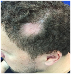
A 47-years-old man presented a localized hair loss in the left temporal region since birth. On dermatological examination was observed triangular alopecia area, well defined, measuring 4 x 3cm (Figure 1) without skin changes, such as atrophy, peeling or inflammation. Dermoscopy (DermLite DL3N; 3Gen) reveled in the injured area vellus hairs, surrounded by normal scalp with terminals hairs (Figure 2). No yellow spots, exclamation mark hairs or cadaver hairs were detected. He denied similar cases in the family.

Figure 1 Well circumscribed triangular alopecia area 4 X 3 cm. No cutaneous alterations such as atrophy, scalling or inflammation.
Case report 2
A 48-years-old male presented a localized hair loss in the right temporoparietal region perceived since she was twelve. On dermatological examination was observed an oval area of alopecia, well-defined, measuring 1 x 1.5cm (Figure 3) without skin changes, such as atrophy, peeling or inflammation. Dermoscopy (DermLite DL3N; 3Gen) reveled in the injured area vellus hairs surrounded by normal scalp with terminals hairs (Figure 4). No yellow spots, exclamation mark hairs or cadaver hairs were detected. He denied similar cases in the family.
The TTA is part of the group of non-scarring alopecia. Unlike the name it was given, most of the cases is not congenital, manifesting from two to nine years old, but may appear during adulthood.2 Remains stable throughout life, is usually unilateral, but about twenty percent are bilateral.3 Often affects the temporal region, but may be located in the temporoparietal region and occipital.4 Some congenital disorders were associated, such as iris nevus syndrome, phacomatosis pigmentovascularis, congenital heart disease, bone and tooth abnormalities, mental retardation and congenital aplasia cutis.2
Histopathology reveals preserved total number of follicles, but most formed by vellus hairs, some intermediaries and lack of terminals. There were no changes in the epidermis and derme.2 The proportional increase of vellus and intermediate hairs in relation to the terminals support the notion that TTA is a form of hamartoma with local follicular morphogenesis alteration.4
Dermoscopy has been an important diagnostic tool, avoiding invasive procedures. TTA most common findings are white hairs, diversity of diameter and normal follicular orifice with vellus hairs surrounded by terminal hairs without atrophy or inflammation.5 It is important to rule out other conditions that preserve vellus hair. Alopecia areata is distinguished by the presence of yellow dots, black dots, exclamation mark hairs, cadaver hairs and dystrophic hairs (Table 1). Trichotillomania shows black dots and traction alopecia is observed the presence of hair casts and cadaverized hairs.6-8
TTA |
AA |
|
Onset |
Congenital or during childhood or adulthood |
Childhood or adulthood |
Location |
Temporal, tempoparietal or occipital |
No site predilection |
Dermoscopy |
Normal follicular orifice with thin vellus hairs, surrounded by terminal hairs at the periphery without atrophy or inflammation |
Yellow dots, black dots, exclamation mark hairs, cadaver hairs and dystrophic hairs |
Histopathology |
Non inflammatory; Preserved total number of follicles, mostly formed by vellus hairs, some intermediaries and lack of terminals |
Peribulbar lymphocytic inflammation, a shift to catagen and telogen follicles and miniaturization |
Table 1 Comparison between TTA and AA.
In 2011, was proposed a classification for the diagnosis of TTA that includes four main features: (I) Triangular or spear-shaped alopecia surrounding the frontotemporal region of the scalp; (II) A normal follicle orifice with vellus hairs surrounded by the terminals (using a dermoscopy); (III) Absence of fractured or exclamation mark hairs, black or yellow dots and absence of a follicular orifice (using a dermoscopy); and (IV) Lack of hair growth 6 months after the confirmation of the presence of vellus hairs, both clinically and dermoscopically.7 Therapeutic options are scarce. One case has been reported in the literature of therapeutic success after using topical minoxidil.9 The treatment of choice is surgery. Depending on the size of the affected area can be attempted excision or, if more extensive, hair transplantation.10 Our patients have chosen not to treat.
Many authors believe that TTA is not an uncommon disorder but that it is under diagnosed due to diagnostic confusion with other types of non-scarring alopecia. It is important to know this diagnosis when facing a patient with non-scarring alopecia and avoid unnecessary treatments.
None.
The authors declared that there are no conflicts of interest.

©2018 Diniz, et al. This is an open access article distributed under the terms of the, which permits unrestricted use, distribution, and build upon your work non-commercially.