Journal of
eISSN: 2373-633X


Case Report Volume 11 Issue 2
Department of Dermatology, University Hospital of Fez, Morocco
Correspondence: Khadija Elboukhari, Department of Dermatology, University Hospital of Fez, Morocco, Tel +212621119073
Received: February 26, 2020 | Published: March 18, 2020
Citation: Elboukhari K, Rasso A, Elloudi S, et al. Senile sebaceous nevus degenerating to a pseudo glandular basal cell carcinoma. J Cancer Prev Curr Res. 2020;11(2):35-36 DOI: 10.15406/jcpcr.2020.11.00422
Jadassohn nevus is a congenital hamartoma that is characterized by natural evolution in three stages, with a risk of malignancies occurring in the last phase. This benign adnexal tumor often affects the face and scalp. Dermoscopy can provide a major help for detecting transforming tumors. We report the case of a senile sebaceous Nevus which has been diagnosed after the occurrence of Basal cell carcinoma in a 60 years old man. The originality of this observation remains in the rarity of the pseudo glandular basal cell carcinoma that, in the best of our knowledge, has never been reported in underlying nevus sebaceous.
Keywords: sebaceous nevus, dermoscopy, basal cell carcinoma
Sebaceous nevus or Jadassohn nevus is a benign adnexal tumor that often occurs in the face and scalp.1 This congenital hamartoma is characterized by a risk of transforming especially in the adolescent stage. Dermoscopy is a non-invasive imaging technique that provides a major help for detecting transforming tumors by giving highly specific features.2 Prophylactic surgery has a large place in the management of this entity.
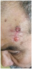
A 60 years old man consulted in our training for a lesion of the forehead evolving for 2 years, occurring on a congenital lesion, this patient had already benefited from his referring doctor, a skin biopsy which revealed a basal cell carcinoma. When examining our patient we noted an infiltrated plaque, containing in its upper part an ulcerated pigmented nodule, in its middle part a scar of biopsy of normal appearance and in its lower part a yellowish plaque, slightly infiltrated, ulcerated by location (Figure 1). When we put our Dermoscopy, we found in the upper area of the lesion, blue-gray ovoid nests with large ulceration and, in the surrounding area, we noted bright yellow spots (Figure 2). We evocated then a basal cell carcinoma or a trichoblastoma in an underlying sebaceous nevus. Our patient has had a total lesion excision total with 0,5mm marges. histopathological examination of the total lesion showed a basaloid cell proliferation with pseudo glandular disposition, adjacent to a hypertrophic sebaceous gland (Figure 3).

Figure 1 Pigmented ulcerated nodule separated from a yellowish translucent plaque by a biopsy scar, located in the left frontal area.
Nevus sebaceous is a complex hamartoma that occurs since birth, and involve the epidermis and dermis, with a triphasic evolution: alopecic patch with underdeveloped adnexal structures. In puberty, the second phase shows a proliferative and veracious plaque. While the third phase is characterized by the occurrence of benign or more rarely invasive tumors in the preexistent plaque.3 Dermoscopy finds an important place in the characterization of these stages of evolution, and especially in the detection of transformation. 10-20% of sebaceous nevus is transformed into benign or malignant tumors that may be epidermal, adnexal or parenchymatous.4,5 This transformation occurs generally in adult life as solitary or several nodules within the confines of the naevus.3 The most common neoplasm occurring in nevus sebaceous is trichoblastoma,3,6,7 followed by basal cell carcinoma,3 those entities present clinic dermoscopical similarities. In fact, in referring to Zaballos et al.3 study Blue-gray ovoid nests, arborizing vessels, and telangiectasis are described in both tumors.3 While other studies attach blue-gray nets to only basal cell carcinoma.2 This dermoscopic features can also be found in hydrocystoma and hidradenoma that occurs rarely in sebaceous nevus.3 Other malignant tumors can occur in this congenital hamartoma, such are Squamous cell carcinoma8 and sebaceous carcinoma.9
On the other hand, in our case we noted a rare subtype of basal cell carcinoma, which is the pseudo glandular variety. To the best of our knowledge, there are no similar cases reported in the literature.
Although the main risk of transformation is in adolescent age, a delayed cancerization may occur especially in the elderly, that justify a regular dermoscopic control of this tumor if a prophylactic surgery has not been conducted.
None.
None.
The authors declare there are no conflicts of interest.

©2020 Elboukhari, et al. This is an open access article distributed under the terms of the, which permits unrestricted use, distribution, and build upon your work non-commercially.