Journal of
eISSN: 2373-633X


Case Report Volume 11 Issue 4
1Senior Consultant Radiologist, Clinical Imaging Department, Hamad Medical Corporation, Qatar
2Assistant Professor in Clinical Radiology, Weil Cornel Medical College, Qatar
3Consultant Radiologist, Clinical Imaging Department, Hamad Medical Corporation, Qatar
Correspondence: Sushila Ladumor, Senior Consultant Radiologist, Clinical Imaging Department, Hamad Medical Corporation, HGH, Doha, Qatar, Assistant Professor in Clinical Radiology, Weil Cornel Medical College, Qatar, Tel +9744 4391249, Fax +974-44392276/1731
Received: August 05, 2020 | Published: August 31, 2020
Citation: Ladumor S, Rashid A. Rare finding in patient with blunt upper abdominal trauma. J Cancer PrevCurr Res. 2020;11(4):95-97. DOI: 10.15406/jcpcr.2020.11.00435
The duodenum is relatively protected due to its retroperitoneal location. An isolated duodenal injury (IDI) involving the duodenum is rarely seen following a blunt abdominal trauma. Here we are reporting rare imaging findings in young patient of 30 yrs. old gentle man presented with blunt upper abdominal trauma with mild upper abdominal tenderness. The main reason to report this case is to identify rare isolated injury in cross-sectional imaging findings, and management of IDI. As IDI can present vaguely and difficult to diagnose clinically. If periduodenal free fluid seen on computed tomography scan and adjacent organs are within normal with no obstructive changes can raise possibility of the duodenal injury. Early diagnosis is very helpful for management as well as important to prevent associated early and late complication.
Keywords: blunt abdominal trauma, duodenal injury, duodenal hematoma, isolated duodenal hematoma
IDI, isolated duodenal injury; CT, computed tomography; MRI, magnetic resonance imaging; EUS, endoscopic ultrasound; ED, emergency department; DPL, diagnostic peritoneal lavage
Our patient who is 30 yrs. old male, had blunt trauma with a heavy metal object that fell on him while lying down and mild upper abdominal pain with tenderness was brought to ED. On examination found to have mild tenderness in upper abdomen and no external sign of trauma. No other complains.
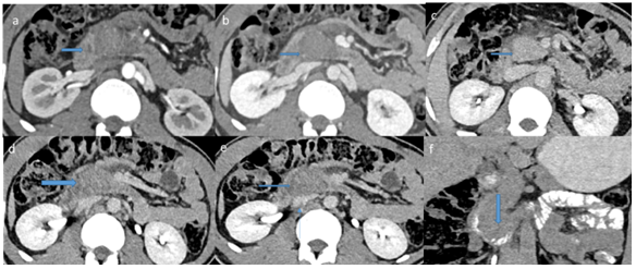
CT scan ordered, done in arterial and venous post-contrast phase which reveal non-enhancing focal lesion at proximal third part of duodenum measured approximately 4x3.5cm (HU=76 in arterial phase) with some fat standing around lesion and is abutting uncinated process of pancreas. No proximal obstruction. Otherwise Pancreas, pancreatic duct and CBD are unremarkable.
No free fluid. No free air. Rest of abdominal and pelvic organs is unremarkable. Oral contrast was given and showed focal luminal narrowing at site of lesion with smooth passage of contrast distally without any signs of obstruction. In view of trauma diagnosed as likely isolated duodenal hematoma, however underlying lesion cannot be excluded Figure 1.

Figure 1 Post-contrast CT scan in arterial (a) venous (b) and delayed (c,d,e) post-contrast phase which reveal non-enhancing focal lesion at proximal third part of duodenum (transverse arrow) measured approximately 4x3.5cm (HU=76 in arterial phase) with some fat standing around lesion and is abutting adjacent head of pancreas(transverse arrow in c).
Post-contrast CT scan in arterial (a) venous (b) and delayed (c,d,e) post-contrast phase which reveal non-enhancing focal lesion at proximal third part of duodenum (transverse arrow) measured approximately 4x3.5cm (HU=76 in arterial phase) with some fat standing around lesion and is abutting adjacent head of pancreas(transverse arrow in c). Later oral contrast was given showing focal luminal narrowing at the site of duodenal lesion (vertical arrow in f) with no proximal obstruction and contrast seen passing distally. Otherwise Pancreas, pancreatic duct and CBD are unremarkable.
(From Cerner). First endoscopy performed and showed stenosis of third part of duodenum. Next day EUS performed revealed circular sub epithelial lesion with intact mucosa in the third part of duodenum. The lesion was inhomogenously hypoechoic and contained liquid part. It is highly suggestive for organizing hematoma. A small part of this lesion is hypoechoic and could be hematoma or sub epithelial lesion.-In comparison to yesterday endoscopy, stenosis of lumen in this part is decreasing.
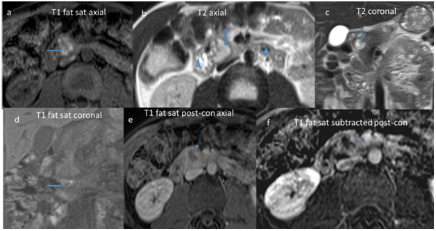
Patient had no obstructive symptoms and other organs were unremarkable so patient managed conservatively. MRI requested after two weeks: Re-demonstration of third part of duodenal lesion which is smaller in size measures about 3x2cm and showing heterogeneous high signal in T1 and T2, diffusion restriction (Dark blood) and no post–contrast enhancement confirmed hematoma which is reduced in size (Figure 2).

Figure 2 MRI requested after two weeks: Re-demonstration of third part of duodenal lesion which is smaller in size measures about 3x2cm and showing heterogeneous high signal in T1 (transverse arrow in a & d) and T2 (vertical arrow in b & c), diffusion restriction (Dark blood, not shown) and no post –contrast enhancement confirmed hematoma which is reduced in size (Thin arrow in e & f). Duodenum proximal and distal to lesion appears within normal (Triangle in b).
Isolated duodenal injury from blunt trauma is not common, occur in less than 2-3% of all abdominal injuries. These type of injuries usually happens as a result of the direct impact on the upper abdomen of the steering wheel or the handlebars during traffic accidents or as direct fall of heavy object on abdomen as in our case.1,2 Diagnosis of a blunt injury of the duodenum only can be challenging as imaging findings are usually subtle. Late diagnosis of isolated duodenal injury may result in complications which range from occlusion or stricture of the duodenum to pancreatitis, hemorrhage, and clinical shock.1–4 The retroperitoneal location of the third part of duodenum, makes physical examination, diagnostic peritoneal lavage (DPL) and ultrasonography becomes relatively less informative in diagnosis of isolated duodenal injury.5
An isolated duodenal injuries after blunt abdominal trauma are not common, however, early diagnosis and treatment can play major role in reducing morbidity and mortality. CT is very important in early detection of duodenal injuries. Large hematoma/injury in view of history of blunt trauma with no other medical history or comorbidity is easy to diagnose with great degree of confidence. Diagnosis of small hematoma with minimal signs of injury with whole-body CT can be difficult. Specific injury patterns in the duodenum often have variable imaging findings at early posttraumatic CT. We have to look very carefully for the subtle signs of injury, like periduedenal fat stranding, duodenal wall thickening, proximal obstructive changes or free air in case of perforation. CT is also helpful and reliably reveals known complication like obstruction, perforation, abscess and extensive injury can have associated pancreatic injury and lead to pancreatitis.1–5
Most patients with intramural duodenal hematoma would respond well to conservative management. If patient develop obstructed over seven to ten days or have evidence of perforation, surgical intervention may be helpful.
Early diagnosis and proper management in cases of isolated duodenal injury is very important to prevent complication and thus mortality and morbidity.
None.
The authors declare there are no conflicts of interest.
None.

©2020 Ladumor, et al. This is an open access article distributed under the terms of the, which permits unrestricted use, distribution, and build upon your work non-commercially.