Journal of
eISSN: 2373-633X


Clinical Images Volume 10 Issue 6
Department of dermatology, University Hospital of Fez, Morocco
Correspondence: Khadija Elboukhari, Department of dermatology, University Hospital of Fez, Morocco
Received: November 21, 2019 | Published: December 10, 2019
Citation: Elboukhari K, Benkirane S, Gallouj S, et al. Pigmented eccrine poroma mimicking a pigmented basal cell carcinoma. J Cancer Prev Curr Res. 2019;10(6):160-161. DOI: 10.15406/jcpcr.2019.10.00410
Eccrine poroma is an uncommon appendage skin neoplasm originating from the terminal distal portion of the sweat glands annexed to the skin. This benign affection commonly appears as a skin colored papules or nodules localized on the extremities. Its pathogenesis may be secondary to trauma scars. We present a case of pigmented eccrine poroma localized on the face, with clinic and dermoscopic appearance similar to basal cell carcinoma.
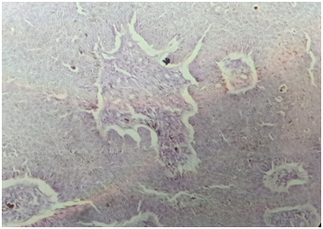
A 38 years old man presented a Black nodule on the face, which appears since childhood and was growing up 02 months before the consultation. It was painless and without pruritus. The dermatological examination showed a sessile-based nodule flesh-colored in places and black in others it was localized under the lower left eyelid. Dermoscopy showed typical ovoid nests and thick arborizing vessels. In front of this clinico dermoscopic panel, pigmented basal cell carcinoma was evoked. Histologic examination showed a proliferation of tumors connected to the epidermis, arranged in masse and made of regular cuboid cells presenting monomorphic nuclei with fine finely nucleated chromatin and an abundant basophilic cytoplasm. Massifs and spans are sometimes centered on cavities filled with eosinophilic material. The tumor cells are pigmented and the mitoses were rare (Figure 1–3).

Figure 3 HES Stain G x 100: cuboid cells proliferation with melanin granules and pigmented melanocytes.
Eccrine poroma is uncommon benign neoplasm first described by Goldman P et al.1 It originates from the terminal ductal portion of the sweat glands annexed to the skin, they commonly appear as a skin colored papules or nodules localized on the extremities.2 Its pathogenesis may be secondary to trauma scars.3 In our patient the nodule localized on the face, which suggest the implication of UV radiations. Although Blue-gray ovoid nests and arborizing vessels are known as a specifics dermoscopy signs of Basal Cell Carcinoma, They have been found recently in pigmented poroma.4 Histologically, the eccrine poroma is recognisable distinct from the other eccrine ductal tumors.5 They are designated as benign lesions due to their lack of cytologic atypia and mitotic activity, as was found in our patient. The most described aspects of pigmented eccrine poroma is the proliferation of uniform cuboid cells with light-colored cytoplasm and evident intercellular bridges.2 We can find also hyperkeratosis, melanin granules and pigmented melanocytes like in our case.
None.
The authors declare there are no conflicts of interest.

©2019 Elboukhari, et al. This is an open access article distributed under the terms of the, which permits unrestricted use, distribution, and build upon your work non-commercially.