Journal of
eISSN: 2373-633X


Short Communication Volume 14 Issue 3
1Department of Clinical Research, Canon Medical Systems, Brazil
2Instituto do Coração do Hospital das Clínicas da Faculdade de Medicina da Universidade de São Paulo (InCor-HCFMUSP), Brazil
Correspondence: Henrique J Cirino, Department of Clinical Research, Canon Medical Systems, Brazil, Tel +55 11 4134-0081
Received: July 14, 2023 | Published: July 25, 2023
Citation: Cirino HJ, Matsumoto JKN, Nomura CH, et al. Impact of silver beam on radiation dose reduction in chest computed tomography: first impressions. J Cancer Prev Curr Res. 2023;14(3):74-75. DOI: 10.15406/jcpcr.2023.14.00524
The continuous technological advances in Computed Tomography have allowed for less exposure to radiation in patients who undergo chest CT, especially in those who control diseases such as cancer. Silver Beam, a beam-shaping energy filter, takes advantage of the photon attenuation properties of silver to selectively remove low-energy photons for low-dose chest examinations. The objective of this study was to evaluate the performance of Silver Beam in dose reduction and image quality in chest CT scans. Acquisition using Silver Beam represented a 8.3 time dose reduction when compared to standard acquisition. Even for patients with large body biotypes, the beam shaping energy filter used in Silver Beam was able to reduce the dose considerably while maintaining the same level of image quality. First impressions of using Silver Beam technology provided high quality CT images, low noise and considerable reduction in radiation dose.
The continuous technological advances in Computed Tomography (CT) have allowed for less exposure to radiation in patients who undergo chest CT, especially in those who control diseases such as cancer, nodules, cystic fibrosis, among others. This may become increasingly important over time to reduce the cumulative radiation dose.1-3
Lung cancer represents a universal public health problem, as it is the leading cause of death from cancer in the world, more than the number of deaths from colon, breast and prostate cancer combined.4 The need to develop other measures against the high prevalence and mortality of lung cancer, in addition to combating smoking, led to the idea of developing screening using imaging methods capable of diagnosing the disease in its early stages.5 Initially, conventional chest X-rays were used, but in the last two decades, with the evolution of radiology as a medical science and the development of increasingly advanced techniques, CT, which allows the study of human anatomy through cross-sectional analysis of the patient through radiation, has gained prominence.6
Silver Beam, a beam-shaping energy filter, takes advantage of the photon attenuation properties of silver to selectively remove low-energy photons from a polychromatic X-ray beam, leaving an energy spectrum optimized for low-dose chest examinations (Figure 1).
Designed to work in combination with the Deep Learning AiCE (Advanced intelligent Clear-IQ Engine) re-constructor, Silver Beam offers enhanced tomographic acquisitions, enabling high quality, low noise and low radiation dose images.
Objective
The objective of this study was to evaluate the performance of Silver Beam in dose reduction and image quality in chest CT scans.
Computed tomography (CT) scans of the chest were performed using a 320-channel CT scanner with 640 slices (Aquilion One Prism-Canon Tokyo, Japan). Helical acquisition, 512x512 matrix, FOV 320–500 mm, slice thickness 0.5 mm, tube rotation 0.5 seconds (standard protocol) and 0.275 seconds (Silver Beam protocol), 120 kV and dose modulation. The images were reconstructed with the Deep Learning Reconstruction Algorithm-AiCE.
Silver Beam provides high quality, low noise CT lung cancer screening images.7 The improvement contained in the algorithm due to artificial intelligence (AI), results in a radiation dose on the order of a typical chest X-ray examination (Figure 2).

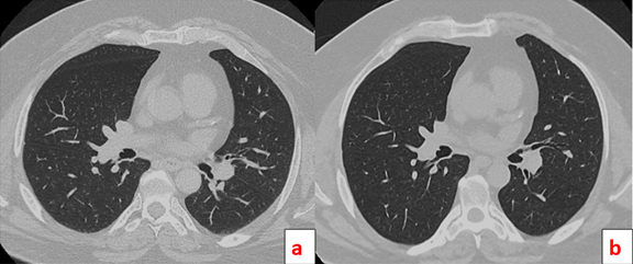
Figure 2 Chest tomography with pulmonary nodule, Image A showing acquisition with standard protocol, Image B using SilverBeam.
The radiation dose of the CTs in figure 2 were respectively: image A CTDIvol = 6.1 mGy/effective dose = 3.10 mSv and image B CTDIvol = 0.6 mGy/effective dose 0.37 mSv. Acquisition using SilverBeam represented a 8.3 time dose reduction when compared to standard acquisition. Due to the use of the reconstructor in Deep Learning AiCE, the image quality remained very similar between the two acquisitions in a patient with a thin biotype.
In a patient with a larger biotype, the same level of dose reduction can be achieved, maintaining excellent image quality (Figure 3).

Figure 3 Patient with large biotype, Image A showing acquisition with normal parameters, Image B using SilverBeam.
Figure 3 shows chest CT images of a patient with a height of 2 meters and a weight of 147 kg. Image A shows the standard acquisition with radiation dose of CTDIvol = 18.30 mGy and, image B, the acquisition performed with SilverBeam with radiation dose of CTDIvol = 2.60 mGy, resulting in respective effective doses of 10.5 mSv and 1.5 mSv. Thus, even for patients with large body biotypes, the beam shaping energy filter used in SilverBeam was able to reduce the dose considerably while maintaining the same level of image quality.
Our study reports the initial results of using a silver filter at the output of a CT scanner's X-ray tube, resulting in a lower radiation dose in chest CT scans. Protocols with lower radiation dose and excellent image quality are increasingly essential in the clinical routine of diagnostic imaging centers. The advancement of technology, combined with the resources of artificial intelligence, has contributed significantly to an accurate and increasingly safe diagnosis for patients. The obtained results allowed verifying an excellent dose reduction between the radiation values obtained from an acquisition with standard parameters, and the acquisition performed with SilverBeam.
In a study that evaluated whether low-dose CT radiation can reduce cancer mortality, Tang et al.,8 showed that a low-dose (twice lower) CT screening for lung cancer was shown to reduce deaths in high-risk smokers. Furthermore, due to its high sensitivity, it has been seen to perform excellently in diagnosing early-stage lung cancer. In our study, it was seen that an even greater reduction (it is worth mentioning that at least 5 times) provided excellent image quality for the detection of pulmonary nodules, with doses lower than 2 mSv.
Another sensitive factor is pediatric patients. Radiation dose is extremely important in children, as they are more prone to developing radiation-induced carcinogenesis due to longer post-exposure life expectancy and more sensitive organs to the oncogenic effects of radiation.9 Dorneles et al.,10 in their study showed that a low-dose chest acquisition can be performed with a submillisievert radiation dose, with preservation of image quality, to allow the identification of lung anatomy, common lung diseases and thoracic neoplasms. The present study evaluated only adult patients, requiring further studies in pediatrics. However, with the levels of radiation reduction seen with the use of SilverBeam, it is expected that ultra-low doses can be achieved while maintaining image quality for this audience.
de Koning et al.,11 in their study also report the cost-benefit ratio in a screening of chest studies using low-dose CT. In addition to saving equipment components, a wide range of scenarios can be contemplated. In this sense, we saw great potential in using SilverBeam in our initial impressions. The ease of automatic integration with the equipment's protocols contributes to a fast and assertive workflow, allowing reports with greater accuracy and efficiency. New studies with more patients and variables should be carried out to investigate the possible outcomes of using silver filters in computed tomography equipment.
First impressions of using SilverBeam technology provided high quality CT images, low noise and considerable reduction in radiation dose when compared to the standard institutional protocol. The obtained results brought relevant perspectives in future studies with optimized protocols.
None.
Authors declare that there is no conflict of interest.

©2023 Cirino, et al. This is an open access article distributed under the terms of the, which permits unrestricted use, distribution, and build upon your work non-commercially.