Journal of
eISSN: 2373-633X


Case Report Volume 7 Issue 6
Department Of Paediatric Hematology and Oncology, The Children's Hospital & The Institute Of Child Health, Pakistan
Correspondence: Arifa Khalid, Fellow Pediatric Hematology/Oncology, Department of Pediatric Hematology/Oncology, The Children's Hospital & ICH, Lahore, Pakistan
Received: February 28, 2017 | Published: March 31, 2017
Citation: Khalid A, Faizan M, Khan S, et al. Extra-renal wilm’s tumor in a 3 years old boy-a case report. J Cancer Prev Curr Res. 2017;7(6):165-166. DOI: 10.15406/jcpcr.2017.07.00254
Wilm’s Tumor, also known as nephroblastoma is mainly a malignant tumor of the kidneys. Extrarenal Wilm’s is a rare entity and only few case reports are available in paediatric literature. We report the case of a 3 years old Pakistani boy who presented with abdominal mass in right upper and lower quadrant associated with pain. Abdominal tomography revealed a huge retroperitoneal mass compressing the right kidney & liver. Trucut biopsy of the mass revealed WT1 positive Wilm’s tumor. He is currently on postoperative chemotherapy and is doing well.
Keywords: wilm’s tumor, extra renal, lumbar mass
Extrarenal Wilm’s tumor (ERWT) is an extremely rare entity with an estimated occurrence of 0.5-1% of all Wilm’s tumor cases.1 It occurs mostly in early childhood; however cases in adults have also been reported. Its location can be retroperitoneal, mediastinal, lumbosacral, inguinal, pelvic, paratesticular and female genital organs as reported in literature.2 This is often misdiagnosed for other common retroperitoneal masses of that region.The exact pathogenesis of extrarenal wilm’s tumor is indefinable until now.3 The clinical presentation is variable depending upon its site and stage. Management and staging of extrarenal nephroblastoma is done according to its renal counterpart.4,5 Hereby we present a rare case of an Extrarenal Wilm’s tumor arising in the retroperitoneum.
A previously healthy, 3 years old boy was admitted in the pediatric Hematology/Oncology department of The Children’s hospital & ICH, Lahore with the complaints of abdominal distension, fever and abdominal pain of 10 days duration. There were no associated vomiting and urinary complaints .The child was normotensive with no clinical dysmorphism. His growth parameters were on the 50th centile.
On examination his abdomen was distended and tense with fullness in the right lumbar region extending upto right hypochondrium. An approximately 10X 8 cm sized non mobile mass was palpable in right hypochondrium and lumbar region. The surface was smooth with well-defined borders and normal overlying skin. The hematological parameters and routine investigation reports were normal. Tumor markers (alpha fetoprotein) was within normal limit.
Abdominal ultrasonography demonstrated a huge heterogenous echogenecity mass extending from the epigastric region into the right hypochondrium and umbilical region abutting the inferior surface of the liver. Right kidney was separately visualized and show mild hydronephrosis. Left kidney was normal.
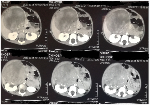
CT scan of the abdomen showed an approximately 20x 10 x 9 cm sized mixed density heterogeneously enhancing mass in the right hemi abdomen. It was compressing and abutting the inferior surface of right lobe of liver pushing it upwards. The mass was also compressing the right kidney and displacing it laterally. There was no abdominal lymphadenopathy. Aorta and IVC appear normal with no vascular encasement [Figure 1 & Figure 2].

Figure 1 CT scan of the abdomen showed an approximately 20x 10 x 9 cm sized mixed density heterogeneously enhancing mass in the right hemi abdomen.

Figure 2 CT scan of the chest showed an atelectatic band in posterior basal segment of left lower lobe with adjacent inflammatory infiltrates.
CT scan of the chest showed an atelectatic band in posterior basal segment of left lower lobe with adjacent inflammatory infiltrates. A tiny suspicious nodule was appreciated in right upper lobe in apical segment. Mediastinal vessels were normal.
Trucut biopsy from the lesion revealed morphologic features of Wilms' tumour with strongly positive immunohistochemistry for WT1.
The child was given preoperative 6 weeks chemotherapy (Vincristine, Dactinomycin, Doxorubicin) according to SIOP WT 2001 protocol.
Re-evaluation CT chest and abdomen showed an approximately 7x5 cm sized mass. There was more than 50% reduction in the size with complete resolution of pulmonary metastasis. Complete resection of the mass was done.
On gross examination the tumor was 7x5x3 cm in size and brown in colour. Histopathological examination revealed Extrarenal Intermediate risk, Regressive type nephroblastoma. There was extensive necrosis, sheets of hemosiderin laden macrophages, sclerosis, fibrosis and areas of calcification. Residual tumor in the form of tubules is also present .morphological features are of post chemotherapy Wilm’s tumor.No focal anaplasia was noted.
Post operatively the CT abdomen showed no residual mass.
Post operative adjuvant chemotherapy was started to the child consisting of Dactinomycin, Doxorubicin and Vincristine and is responding well to treatment.
Wilm’s tumor is the most common pediatric malignancy of renal origin. Extra renal Wilm’s tumor is very rare. Its clinical presentation depends upon the location and stage of the tumor. The most common sites of ERWT are retroperitoneum and inguinal canal as per literature review. Retroperitoneal ERWT are more common in males.6,7 Similar finding is observed in our case. The exact mechanism whereby a Wilm’s tumor occurs in extrarenal tissues is unkown. However, according to a popular hypothesis they arise from ectopic metanephric blastema.8
The imaging features of extrarenal wilm’s tumor are nonspecific and the heterogeneous appearance as defined in the earlier reports was also encountered in our case. Therefore, the diagnosis of ERWT proposed on imaging studies can be confirmed only upon its operative removal and pathological evaluation of the specimen.9 The staging system used in the literature while describing the case was NWTS (National Wilm’s Tumor Study) system. The prognosis of ERWT when matched with the appropriate stage is similar to its renal counterparts.10
ERWT is a rare malignant neoplasm with atypical presentations. This report suggests that ERWT should be considered in the differential diagnosis of the retroperitoneal tumors. The staging, response to chemotherapy and prognosis are similar to the renal Wilm’s counterpart. Therefore, similar staging and treatment protocol can be followed.
None.
None.
The authors declare that there is no conflict of interest.

©2017 Khalid, et al. This is an open access article distributed under the terms of the, which permits unrestricted use, distribution, and build upon your work non-commercially.