Journal of
eISSN: 2373-633X


Case Report Volume 5 Issue 1
Department of Radiology, University of Minnesota, USA
Correspondence: Hamed Jalaeian, Department of Radiology, University of Minnesota, 420 Delaware Street SE, MMC 292 , B-212 Mayo, Minneapolis, MN 55455, USA, Tel 612.626.5589, Fax 612.624.3188
Received: April 14, 2016 | Published: June 7, 2016
Citation: Jalaeian H, Taylor AJ. Accidental foreign body ingestion resulting in detection of an asymptomatic colorectal carcinoma. J Cancer Prev Curr Res. 2016;5(1):206-207. DOI: 10.15406/jcpcr.2016.05.00148
Foreign body ingestion is a fairly common clinical problem. The clinical course and resultant management is typically uncomplicated and will usually be predicted from the characteristics of the foreign body itself. However, an abnormal gastrointestinal tract segment can result in a complicated outcome. This article demonstrates such an example when an ingested foreign body that should have passed uneventfully, instead caused colonic obstruction by obturating a previously asymptomatic colon carcinoma. A 70 year old gentleman presented to the emergency department with onset of abdominal pain a few days following accidental ingestion of his dental crown. An initial plain abdominal radiograph followed immediately by a contrast enhanced abdominal MDCT showed an intraluminal metallic density foreign body, compatible with the ingested crown, causing obstruction at the splenic flexure lodging at a 3cm focal “apple core” lesion. Rapid decompression via expedited colonoscopy and placement of a colonic stent was then facilitated. Acute colonic obstruction due to accidental ingestion of a foreign body is rare. Plain film radiography may suffice for imaging of the uncomplicated foreign body ingestion. However, CT scanning, with the added technical development of 3D reconstruction capability can provide critical information in certain circumstances.
Keywords: ingested foreign body, large bowel obstruction, plain radiography, CT
The adult presenting with possible ingested foreign body is a fairly common event. Most of these ingested objects should pass without difficulty or intervention.1,2 Even so, imaging will be needed; first, usually plain radiography, to define the presence, morphology, and location of the foreign body. At times follow up plain radiography will be required to monitor foreign body passage. But imaging, especially CT, may be critical in this patient population if the foreign body is not very radiopaque or if a complication is questioned. The present case illustrates the importance of rapid imaging for what should have been an otherwise uneventful passage of a foreign body whose impaction at the splenic flexure uncovered an asymptomatic colon cancer.
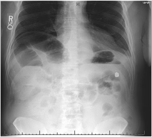
A 70 year old gentleman presented to the emergency department with progressive, intermittent abdominal pain and bloating of 4-5 days duration, associated with nausea, vomiting, and hiccups. He also reported a recent change in his usual bowel habits and a 20-pound weight loss over the prior 6 months. His last screening colonoscopy, 10 years ago, revealed only benign hyperplastic colonic polyps. On a recent visit to his primary care physician, his fecal occult blood test was found to be positive and was scheduled to repeat his colonoscopy on a nonemergent basis. There was no family history of colorectal cancer. On physical examination, his vital signs were stable. His abdomen was distended and tender to palpation, mainly in the epigastric region. An abdominal series (Figure 1) revealed distended ascending and transverse colonic bowel loops along with distended small bowel and numerous air fluid levels all suggesting a colonic obstruction. In addition, a 1.6×1.2cm, fairly smooth, radiopaque object was visualized in the area of splenic flexure. On further questioning, following the abdominal series, the patient recalled accidentally swallowing an old dental crown a few days prior to this presentation. An immediate contrast enhanced MDCT of the abdomen and pelvis was then obtained. The metallic density intraluminal foreign body was confirmed to be at the splenic flexure immediately proximal to a thickened segment of colon. There was distended bowel proximal to this complex and decompressed colon distally Figure 2(A). Better displayed on multiplanar reformatting of the MDCT source images, a 3cm focal area of circumferential mural wall thickening moderately encroaching on the lumen was found at the splenic flexure with an appearance of an “apple core” lesion Figure 2(B). There were adjacent multiple small pericolonic and mesenteric lymph nodes. The gastroenterology service was consulted for urgent colonoscopy and large bowel decompression. On colonoscopy, a fungating, circumferential large mass lesion was found at the splenic flexure. The metallic foreign body, corresponding to patient’s the dental crown, was found immediately proximal to the mass lesion (Figure 3). Biopsies were taken from the colonic mass lesion and at this same setting a colonic uncovered 10cm×25mm×30mm flanged stent was placed. Pathologic examination of the tumor biopsies was consistent with moderately differentiated invasive adenocarcinoma. Patient underwent laparoscopic-assisted subtotal abdominal colectomy with an ileosigmoid anastomosis 2 weeks later. A total of 46 biopsied lymph nodes were negative for malignancy and the patient referred to oncology service for further cancer planning and treatment.

Figure 1 Upright abdominal radiograph demonstrates a metallic foreign body in the left upper quadrant. In addition, there is both distended colon coursing back from the area of the splenic flexure as well as dilated small bowels loops all with air fluid levels. These findings point toward a colonic obstruction with the origin near the splenic flexure.
Most foreign body ingestion in the adult is accidental and usually related to some mental impairment, being edentulous, or having dentures. However, prisoners seeking secondary gain and drug smugglers will intentionally swallow foreign bodies. In general, the ingested material has a high likelihood of passage without significant intervention. Even so, the initial clinical management requires definition of the foreign body’s physical characteristics and position3 usually obtained by imaging. If the foreign body is very likely radiopaque, per history, plain radiographs can be used to assess the foreign body(s) appearance and also confirm its initial position. Subsequent radiographs can then be judiciously used to follow the object’s travel and document its eventual elimination. CT may be necessary if the foreign body is less radiopaque (fish bones, chicken bones, thin metal or aluminum objects, plastic, or wood) or if there is question of possible complication (gastrointestinal tract impaction, obstruction or perforation).3 The addition of 3D rendering with CT can be very helpful.4 A foreign body with a diameter of 2-2.5cm may be stopped in either the esophagus or by the pylorus.3,5 An object greater than 5 cm in length can be halted in the stomach, duodenal sweep, or the ligament of Trietz.5 If a foreign body traverses these normal anatomic narrowings, the ileocecal valve may be a final hurdle to passage.
But additional abnormalities can also cause a foreign body stasis: esophageal dysmotility; gastrointestinal tract nonanatomic narrowings caused by inflammatory/infectious strictures, intramural or perienteral neoplastic processes, or adhesions. The transit of ingested foreign body may reveal these problems. The present case illustrates this last point with what should have been an innocuous foreign body uncovering what will hopefully be a curable, partially obstructing colon carcinoma.
While most ingested foreign bodies will pass without difficulty, imaging is required. Relatively radiolucent foreign bodies, stasis at an unusual site, or clinical suspicion of complication should prompt the use of CT for optimum display and appropriate management.
The authors do not declare any conflicts of financial interests with respect to this manuscript. Both authors have contributed to the collection of data about the case, conception, interpretation of the case and imaging findings, drafting of the article and final approval of the article and meet the criteria for authorship.
None.

©2016 Jalaeian, et al. This is an open access article distributed under the terms of the, which permits unrestricted use, distribution, and build upon your work non-commercially.