Journal of
eISSN: 2473-0831


Research Article Volume 8 Issue 2
1Department of Zoology, Ranchi University, India
2Department of Zoology, St. Xavier’s College, India
Correspondence: Rakesh Ranjan, Department of Zoology, Ranchi University, Ranchi-834008, Jharkhand, India
Received: February 26, 2019 | Published: April 25, 2019
Citation: Ranjan R, Dandapat S, Kumar M, et al. Synthesis and characterization of Cuscutareflexa (Roxb.) aqueous extract mediated silver nanoparticles. J Anal Pharm Res. 2019;8(2):80?83. DOI: 10.15406/japlr.2019.08.00317
C. reflexa is a parasitic medicinal plant has been used for ailment of different diseases. Aqueous extract of C. reflexa has been used for the synthesis of SNPs .In present work synthesis and characterization of SNPs mediated by C. reflexa aqueous extract has been done. The synthesized SNPs showed SRP at 442.94nm. SEM image analysis provided the spherical shape of the SNPs. DLS analysis provided the synthesized SNPs were 169.10nm in diameter with -23mV zeta potential. FTIR spectroscopy provided the confirmation about synthesis of SNPs by representing various transmission peaks 3062.96cm-1 represented to O-H stretch for alcohol and phenol, 2735.06cm-1, 2395.59cm-1, 2063.83cm-1 represented to N-H stretches for primary and secondary amines, 825.53cm-1 for C-H stretch represented to vinyl group. Thus, C. reflexa extract can be used for synthesis of metallic nanoparticles for pharmacological studies.
Keywords: nanoparticles, characterization, shape, number
NPs, nanoparticles; NSMs, nanostructured materials; AgNPs/SNPs, silver nanoparticles; UV-vis, ultraviolet visible spectroscopy; SEM, scanning electron microscopy; DLS, dynamic light scattering; FTIR, furior transform infrared
Nanoparticles (NPs) and nanostructured materials (NSMs) represent an active area of research with full expansion in many applications such as engineering, electronics, pharmaceuticals etc. NPs and NSMs have gained prominence in technological advancements due to their marvellous physicochemical characteristics such as melting point, wettability, electrical and thermal conductivity, catalytic activity, great adsorption capacity with ligands, stability in biological solution, light absorption and scattering resulting in enhanced performance over their bulk counterparts.1,2
But the principle of nanotechnology and it applications with pharmacology and medicine are different and they concern with NMs of diameter in the range of 1 to 100nm. In terms of pharmacology NPs and NSMs are defined as particulate dispersions or solid particles drug carrier and have the property to entrap, encapsulate or attach or insert the drug molecules into a nanoparticle matrix or insert the drug molecules in the core of nanoparticles.3,4
Recently, synthesis of different metallic nanomaterials such as copper, zinc, titanium, magnesium, gold, alginate and silver mediated by biological agents such as bacteria, actinomycetes, fungi, algae, extracts from plant and animal materials etc. have been used for eco friendly and green technology.5,6 Cuscuta reflexa Roxb. Belongs to family Cuscutaceae a division of Convolvulaceae commonly known as Dodder or Akash bel is a parasitic climber plant without leaves has been used as traditional medicine in Ayurveda for treatment of eye and heart and other diseases for a long time.7,8 It has also been reported C. reflexa contains different types of phytochemicals such as, cuscutin, amarbelin, betasterol, stigmasterol, kaempferol, dulcitol, myricetin, qurecetin, coumarin and oleanolic acid which are associated with antispasmodic, heamodynamic, bradycardia, antisteroidogenic, antihypertensive, antiviral and anticonvulsant activities.9,10
The aim of present study dealt with the synthesis of silver nanoparticles using aqueous extract of C. reflexa and their characterization.
Collection of plant material and preparation of extract
Fresh stems of C. reflexa were collected and brought to Department of Zoology, Ranchi University, Ranchi. Fresh stems of C. reflexa was washed by distilled water and then by absolute ethyl alcohol (99.8%) to avoid microbial contamination. The stems were dried in shade under room temperature, powdered and sieved. 50g of the fine powder was subjected to extractraction chamber of Soxhlet and 300mL distilled water was taken in boiling flask as extraction solvent for aqueous extraction. The extract obtained was filtered, concentrated and dried in rotary flask evaporator maintained at 45ºC for proper dehydration and the dried extracts were stored in air tight containers at room temperature for further studies.11
Synthesis of nanoparticles
The synthesis of silver nanoparticles was done with slight modification of previous method of Dandapat et al.12 Synthesis of nanoparticles were done by mixed 3mL (41mg/mL) of C. reflexa aqueous extract and 197mL of 0.1M silver nitrate (169.87g/mol) solution (i.e., 3.35g AgNO3/197mL of distilled water) and incubated by using hot plate at 80ºC and continuous stirring using magnetic stirrer bar, until the light yellow colour of the solution was changed to dark brown. Then the solution was cooled to room temperature and centrifuged at 15000rpm for 10 minutes. The supernatant was discarded and the pellet was washed with distilled water and was dried in the incubator at room temperature for characterization.
Characterization of nanoparticles
Characterization of synthesized silver nanoparticles were done by UV-vis spectroscopy, SEM photography, DLS analysis and FTIR analysis. UV-Visible spectra analysis was done by using Parkin Elmer Lambda-25 UV-Visible spectrophotometer. SEM was done using JEOL JSM-6390 LV (Japan) machine provided with Vega TC software. The DLS analysis for particle size was carried out using Malvern Nano ZS green badge) ZEN3500 (U.K.) zetasizer provided with zetasizer Nano software. FTIR spectroscopy analysis was carried out by IPRresting-21 (Shimadzu Corp., Kyoto, Japan).6,12
Synthesis of nanoparticles and UV-Vis spectroscopy analysis
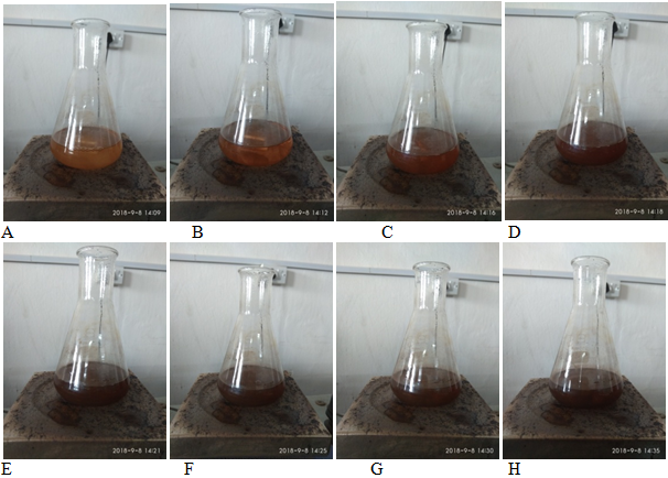
Synthesis of silver nanoparticles mediated by aqueous extract solution is presented in Figure 1. Result showed the change of pale yellow colour of mixed solution of extract and AgNO3 solution turns into dark brown as the temperature and incubation time period increase, which indicates the formation of nanoparticles. Previous work also reported the transformation of pale yellow colour of mixed silver nitrate and Cardiospermum halicacabum L. leaf extract changed to dark brown colour with increase in incubation temperature and time.13 Similar work also preformen previously and reported that also formed dark brown colour from pale yellow colour when Eclipta alba leaf extract and AgNO3 solution were incubated with increasing time.14 Gradual colour change of nanoparticles sample from light to dark colour is an indication of formation of nanoparticles by reduction of metallic silver ion (Ag+) into Ag0 to form AgNPs.15 It has been reported that metabolites of plant extract trapped on the Ag+ ions due to electrostatic interactions between the silver ions and metabolites in plant extract and form their secondary structural change (Ag0) and further reduction of silver nuclei turns in to AgNPs by stabilizing activity of phytochemicals.16

Figure 1(A-H) Colour transformation of mixed C. reflexa and silver nitrate solution with increasing time period and temperature.
UV-vis spectroscopy is one of the most commonly used technique for the characterization, stability, morphology, shape, size, and chemical surroundings of synthesized nanoparticles and it provides surface plasmon resonance (SPR) band due to the combined oscillation of electrons on valence band and conduction band on the surface of AgNPs.17,18 In previous study UV-vis spectroscopy of Costus pictus extract mediated nanoparticles provided adsorption peak at 420nm.19 Previously UV-vis analysis of C. mathewii extract mediated water suspended AgNPs studied and reported a peak of absorption at 423nm that suggested the presence of AgNPs due to surface plasmon resonance.20 The absorption spectrum of nanoparticles obtained from UV-visible absorption spectroscopy is presented in Figure 2, which shows peak at 442.94nm corresponds to the SPR and correlate with the respective UV-Vis spectra of the colloidal solution SNPs of previous studies.
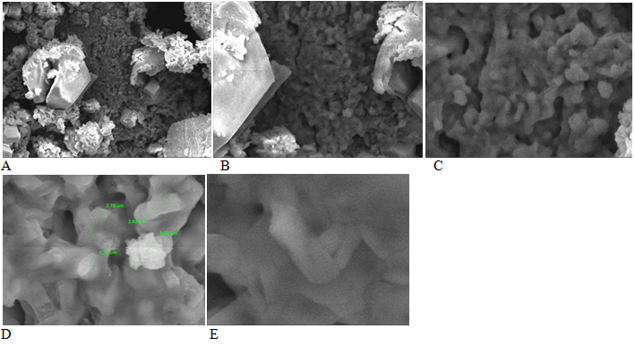
SEM is a surface imaging technique and resolves various molecule sizes, size distributions, nanomaterials shapes, and the surface morphology of the particles at the small scale and nanoscales range.21 Results of SEM images analysis of C. reflexa extract mediated silver nanoparticles (Figure 3) showed that the synthesized nanoparticles were spherical and smooth surface shaped. SEM image analysis provided very large sized nanoparticles of 1.78µm to 2.68µm diameters. Large size nanoparticles were formed due to agglomeration of nanoparticles on drying for SEM analysis of C. reflexa extract mediated SNPs powder. It was reported that Costuspictus extract mediated SNPs were spherical in shape, with an average size of around 100nm and uniformly distributed silver nanoparticles on the surface of the cells was observed.19 Present study correlates with the SEM analysis of SNPs of previous studies.

Figure 3 SEM photographs of C. reflexa extract mediated silver nanoparticles (A: 1000X, B: 2500X, C: 5000X, D: 10000X, E: 20000X).
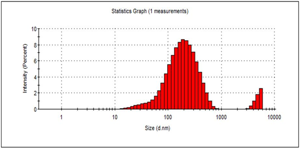
DLS Analysis provides nanoparticles size distribution size and zeta potential, which were important characterizations of NPs because they govern the size distribution by intensity, volume, number and saturation solubility of NPs in colloidal solution and even biological performances.22,23 Result of DLS analysis C. reflexa extract mediated silver nanoparticles is presented in Figure 4-6.

Figure 4 DLS analysis of C. reflexa extract mediated silver nanoparticles by intensity % distribution.
The result showed silver nanoparticles of 15.69nm to 105.7nm diameter were distributed by intensity. But nanoparticles of more than 100nm diameter were formed during synthesis. The DLS analysis provided average 169.10nm diameter silver nanoparticles were distributed in 1mL of nanoparticles sample solution by intensity. However, the 19.05nm of SNPs were distributed with 100 % number distribution. SNPs of 42.43nm and 346.7nm were distributed with 57% and 16 % respectively by % volume distribution. Zeta potential measures the effective electric charge on the nanoparticle surface and provides characterization parameter for stability in aqueous AgNPs suspensions.24 In present study C. reflexa extract mediated SNPs showed -23mV presented in Figure 7. It has been reported that SNPs with zeat potential -25mV to -+25mV posses good stability in aqueous suspension.25 DLS analysis SNPs of present study correlates with the DLS analysis of SNPs of previous studies.
FTIR analysis deals with the probable mechanism behind the formation of SNPs and provides the information about functional groups present on the surface on SNPs.12,26 Result of infrared spectrum analysis of C. reflexa is presented in Figure 6. The result reveled absorption peaks 3062.96cm-1 represented to O-H stretch for alcohol and phenol, 2735.06cm-1, 2395.59cm-1, 2063.83cm-1 represented to N-H stretches for primary and secondary amines, 1708.93cm-1 represented to C=O for conjugated aldehydes and acids, 1570.06cm-1 and 1508.33cm-1 represented to C=C stretches for aromatic compounds, 1384.89cm-1 represented to NO2 and S=O stretches for sulphonyle chloride and sulphonamides, 1072.42cm-1 and 1049.28cm-1 represented to C-N stretch for amines, 825.53cm-1 for C-H stretch represented to vinyl group and 729.09cm-1 and 540.07cm-1 for halo alkans (Figure 8).27-29
Present study concludes C. reflexa extract can be used for synthesis of metallic nanoparticles. The extract is useful for synthesis of stable SNPs and can be used further various pharmacological and medical application without producing any toxic substance and pollutant.
None.
The author declares that there is no conflicts of interest.

©2019 Ranjan, et al. This is an open access article distributed under the terms of the, which permits unrestricted use, distribution, and build upon your work non-commercially.