Journal of
eISSN: 2473-0831


Research Article Volume 6 Issue 3
Correspondence: Morteza Sasani Ghamsari, Photonics & Quantum Technologies Research School, Nuclear Science and Technology Research Institute, North Karegar, Tehran, Iran, Tel -88221005
Received: October 26, 2017 | Published: November 5, 2017
Citation: Razzaghi D, Ghamsari SM (2017) Graphene Plasmonics: A Powerful Sensor and Pharmaceutical Analytical Tool. J Anal Pharm Res 6(3): 00179. DOI: 10.15406/japlr.2017.06.00179
Sensors and analytical tools are very important instruments that play critical role in pharmaceutical research activities related to pharmaceutical sciences. Over the last decade these instruments are largely modified all the time by the development of R&D and innovation output. To make a high-resolution and accurate-mass instrument a novel spectroscopy technique such as Mid-infrared (IR) absorption spectroscopy and surface plasmonic resonance (SPR) were developed for bio-chemical sensing and analytical characterization. In this study, we reviewed the key distinguishing features of graphene plasmons and highlight its potential to b eemployed as pharmaceutical sensor and analysis tool. Finally, we discuss the new opportunities for graphene plasmonics in pharmaceutical manufacturing plants.
Keywords: graphene plasmonics, sensor, analysis tool, pharmaceutical research
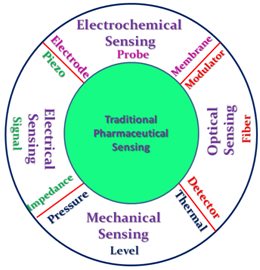
During the last decade, the development of R&D and innovation activities lead to large modifications in analytical tools and sensors that are capable of real-time process monitoring and providing specific production information. Especially, in pharmaceutical and biotech industries, different types of sensors such as temperature, humidity, pressure and membrane sensors are used in different processes in pharmaceuticals. On the other hand biomedical sensors are demonstrated as powerful instruments to be employed in the cancer detection, innovative drugs, new targets and better efficacy/safety requirements.1-3 As shown in Figure 1, different types of sensors and traditional pharmaceutical sensing procedures are used in pharmaceutical industry.

Figure 1 Traditional pharmaceutical sensing procedures and different kinds of sensor used in pharmaceutical industry.
Usually, sensor accuracy and resolution are important parameters that need to be carefully considered for process control and environmental monitoring. In many pharmaceutical industries a supervisor in charge has responsibility to record the pharmaceutical sensed information manually. This kind of data recording process is often involved in data entry errors that need to be addressed. Consequently, real time capturing of the pharmaceutical information and process data is necessary.4 Therefore, advanced track and trace techniques must be developed which so far has not been implemented in the majority of pharmaceutical sensing and analysis methods. It means that we have to specifically focus on identifying the novel sensor device and sensing technology which can be implemented in pharmaceutical manufacturing plants. Recently, optical and wireless sensors are developed as new implements in pharmaceutical manufacturing plants.5 Moreover, more progress must be achieved to assure that new sensor technology can satisfy all requirements of data collection for pharmaceutical manufacturing plants.
In the analysis and testing stages that play an important role in pharmaceutical manufacturing, we involve with the characterization of raw materials, intermediate products and finished dosage forms. As mentioned above, these are needed to achieve some advances in analytical testing tools that can help us to address challenges in pharmaceutical analysis. During the past decade, the significant rise in the small-molecule generic drug, continuous manufacture (CM) and orthogonal recognition of the complex nature of the proteins are three key trends have largely been received in the development of pharmaceutical analytical instruments.6 The first development provides an increase in the instrument accuracy that offers performance-relevant measurements and/or high informational productivity for deformulation and the demonstration of bioequivalence (BE). The second one leads to the scale-up ability of the continuous processes and the ability to file on the basis of full-scale experimental data monitoring and automated control.6 Nevertheless, significant progress that has been made to complement traditional techniques with newer/less well-established analytical tools, we need to build up instruments with higher resolution and accuracy to maximize the collected analytical data. For instance, to learn more about small molecules and biologics, we have to use more sophisticated, specific, and in some cases, more expensive tools. To make a high- resolution and accurate-mass instrument a novel spectroscopy needs to be used for the identification of the biochemical building blocks of life, such as proteins, lipids, and DNA by accessing their vibrational fingerprints.7 Mid-infrared (IR) absorption spectroscopy is one of the powerful techniques that can be used for bio-chemical sensing and analytical characterization. However, the large mismatch between mid-IR wavelengths (2e6mm) and biomolecular dimensions (<10nm) limits the probability of interaction between mid-IR light and nanoscale size biomolecules.8 Over the last decade, a profusion of approaches such as surface enhanced infrared absorption (SEIRA) technique have been made to find solutions to this limitation.8 It seems that the incorporation of various nanomaterials in the construction of sensors and biosensors leads to considerable improvements in their performance and help us to overcome the current technical challenges.9 As a two dimensional (2-D) nanomaterial with ultrahigh carrier mobility, graphene can be used as a plasmon waves host that exhibit extremely tight spatial confinement, exceptionally long plasmon lifetime, and an electrostatically tunable response in the mid-infrared (mid-IR) and terahertz (THz).10 These characters render graphene a viable plasmonic material for achieving novel functionalities in various mid-IR to THz photonic systems. In this paper, we reviewed the key distinguishing features of graphene plasmons and highlight its potential to be employed as pharmaceutical sensor and analytical tool. In addition, the graphene-based sensor ability to monitor the progress of chemical reactions in situ and the ability to determine the cleanliness of pharmaceutical equipment surfaces without the need for swabbing and off-line analysis of the swab will be covered in this column. Finally, we discuss future challenges and new opportunities for graphene plasmonics in pharmaceutical manufacturing plants.
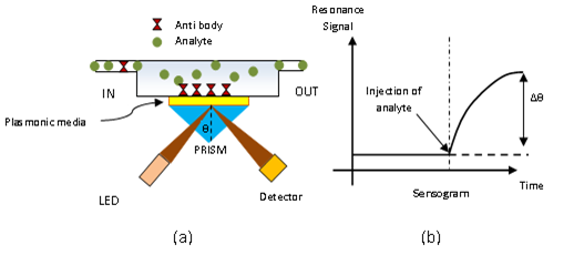
Surface Plasmon Resonance and mid-infrared spectroscopy are among the promising techniques in the field of pharmaceutical analysis and processes.9,10 For example, SPR biosensors, having the important benefit of completely label-free measurement, were successfully applied for a wide variety of (biological) molecules with different sizes, such as small molecules, Mr < 500 Da,11 to larger and more complex structures, including proteins, nucleotides, viruses, and peptides with therapeutic potential.12-15 At the same time, the method provides comprehensive information to: (a) identify the binding typology of a pair of reactants (or more) as a function of the other interacting partner, (b) determine the affinity constants related to the interaction processes occurring during the real-time analysis, (c) quantify association and dissociation rates, and (d) determine the concentration of one/both of the interacting partners. To clarify the outstanding features of graphene and graphene-based plasmonic as sensor and pharmaceutical analytical tools, a brief discussion is made about SPR biosensors which are widely used in pharmaceutical analysis.16 The basis of SPR biosensor is generation of evanescent electromagnetic waves in interface of the thin metallic layer (for example via total internal refraction) which penetrate the thin metal film and tunnel through it to excite surface plasmons at other metal dielectric interface. Under certain angle of light incidence, resonance occurs and the light energy is efficiently transferred to surface plasmons so that a significant drop in intensity will occur. The resonance angle is very sensitive to a change in the refractive index of the dielectric (sensed medium) which is the key to biosensing via SPR phenomenon. It is worth mentioning that phase matching condition which is the perquisite for SPR, can be accomplished by several ways such as using proper grating or waveguide as is discussed by Wijaya et al.17 Many sensor chips have been developed using gold as the plasmonic media. However, gold based sensors suffer from high loss (especially in infrared region) and lack of tunability of the gold thin film.18 High loss, limits the sensitivity and lack of tunability limits the diversity of sensible bimolecules.
Due to these drawbacks of metal plasmonics, it is desirable to use a new plasmonic material to boost the sensors efficiency. Fortunately graphene was one of the suitable candidates as will be discussed here. Graphene is a single-atom-thick planar sheet of sp2-bonded carbon atoms perfectly arranged in a honeycomb lattice.19 Graphene exhibits relativistic like linear energy dispersion and unlike metals its absorption is only depended on the fine structure constant.20-23 Plasmon in graphene originates from the collective motion of massless Dirac fermions, and the carrier density dependence is different from conventional plasmons. Graphene plasmons strongly couple to molecular vibrations of the adsorbents, polar phonons of the substrate, and lattice vibrations of other atomically thin layers.24 Because of its two-dimensional nature, free carrier can be induced through electrical gating or chemical doping which is the reason of optical tuneability of graphene. These properties make graphene a powerful material for sensing and spectroscopy. The plasmonic resonance of graphene matches to mid-infrared to terahertz region so that graphene has found its application in information and communication, medical sciences, homeland security, military, chemical and biological sensing, and spectroscopy.25 Graphene also exhibits extraordinary physical and chemical characters that make it as an exciting new material with wide range of applications including transparent conductors and electrodes in nanoelectronic devices, solar cell, water splitting and organic light emitting diode.26 It has also been used in field emission display.27 In summary three main merits exists for graphene as a plasmonic material. The first one is the high mobility of the carrier in graphene (low loss plasmonic material). The second one is the tunability of the fermi level via electrostatic gating or chemical doping and third one is the very strong light confinement due to the small wavelength of the graphene plasmon compared with light wavelength. In terms of sensing technology, higher resolution compared with traditional sensors is expected from graphene because of its small spatial extension compared with the light wavelength. Also, higher sensitivity is achieved with graphene because of its strong interaction with light over a wide range of frequencies. Interestingly, the chemical potential in graphene can be tuned using an external fields, making the control of light–graphene interaction feasible so that scanning the absorption frequencies of biomolecules, for example, is simply feasible with changing the external field.23,28,29 Beside infrared and mid infrared region, by enhancing the carrier density in graphene, stronger light–graphene interaction in the terahertz and far-infrared ranges will be achieved.29 For example, Fu et al. analyzed the transmission of THz waves through graphene layers and found that the strong attenuation and an enhanced absorption of the THz wave takes place, which is shown to be rooted in the increase of the doping level in graphene, the accumulation of the photo-induced carrier at the interface of the graphene as well as the scattering between carriers, phonons and defects.30 The results of this study imply promising applications for the development of high-performance THz modulators and absorbers.30 In Figure 2, different applications of graphene in the sensing technology are schematically shown (Figure 3).

Figure 2 (a) Schematic of a SPR based sensor in which total internal reflection is used for phase matching, and (b) the resonance signal vs time before and after the injection of analyte.
Liu et al.31 have critically and comprehensively reviewed the emerging graphene-based electrochemical sensors, electronic sensors, optical sensors, and nanopore sensors for biological or chemical detection. Ang et al.16 have successfully demonstrated the solution-gating of epitaxial graphene where ambipolar characteristics with a narrow p-nplateau region (∼0.2 eV) near the Dirac point is observed. They have found that both nand pcarriers induced by capacitive charging of the ideally polarizable graphene/electrolyte interface, with the negative gated potential region exhibiting supra-Nernstian response to pH. Their obtained results showed that its possible applications in solution-gated, ultrafast, ultralow noise biosensors or chemical sensors due to the sensitive response of graphene to surface charge or ion density.32 Purkayastha et al.33 proposed a surface plasmon resonance (SPR) based gas sensor in terahertz frequency with Otto con-figuration based on attenuated total reflection (ATR) technique using free standing doped graphene monolayer. They have investigated the different chemical potential of graphene monolayer to study its effect on these performance parameters.33 Their obtained results showed that the optimization of the gap distance between the prism base and the graphene monolayer has significant effect on the reflectivity of SPR sensing. Li et al.34 reported a novel sandwich biosensor for detection of thrombin using electrochemiluminescence method based on dual-signal amplification strategy that can be successfully applied to thrombin analysis in diluted human serum samples. This biosensor also showed good selectivity for thrombin with-out being affected by some other proteins, such as BSA and lysozyme and so on.34 Graphene nanosheet can also be used as gas sensor. Liu et al.35 developed a flexible, simple-preparation, and low-cost graphene-silk pressure sensor based on soft silk substrate through thermal reduction was demonstrated. They used silk as the support body and provided a three-dimensional structure with ordered multi-layer structure as device format. They found that, graphene-silk pressure sensor can achieve the sensitivity value of 0.4 kPa-1, and the measurement range can be as high as 140kPa.35
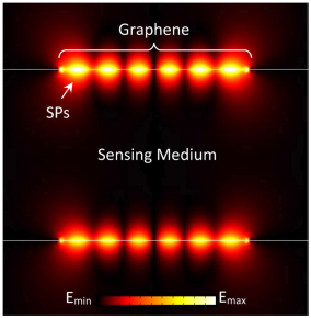
Electrochemical sensing is one of the important applications of graphene-based sensors. As well known as, in electrochemical sensors, a transduced biological or chemical signal is converted into an electrical signal by an electrode. According to the converting technique used to collect the electrical signal is measured as potential, conductivity and current respectively. These utilization techniques provide ability for fast, simple and direct analysis in an even turbid sample matrix. Therefore, electrochemical sensors hold a leading position among chemical sensors.36 Due to the high surface-to-volume ratio and therefore, strongly adsorption of biomolecules to the surface of graphene, it can be used to recognize the properties of biochemical molecular such as antibody-antigen binding and enzymatic reactions employed for selective analysis. Recently a lot of papers have been published in this regard.37-41 Cinti et al.42 provided a critical perspective related to advantages and disadvantages of using graphene in biosensing tools, based on screen-printed sensors. They concluded that graphene can be considered an outstanding polyedric material, since it can be used to tailor the electrochemical properties of a screen-printed electrode, as well as act as a label and loading agent for biomolecules and inorganic nanomaterials, exploiting its high surface area and easy functionalization.42 Bo et al.43 showed that the alleviation or the minimization of the aggregation level for graphene sheets would facilitate the exposure of active sites on graphene and effectively upgrade the performance of graphene-based electrochemical sensors and biosensors. Their founding confirmed that the less aggregated graphene with low aggregation and high dispersed structure can be used in improving the electrochemical activity of graphene-based sensors.43 Recently, Chorsi et al.44 designed a label-free, graphene based biosensing device with ultrahigh sensitivity that operates in the IR frequency band.44 Their device can achieve a sensitivity of up to 3053nm/refractive index unit (RIU), which is three times greater than the sensitivity of current top-level, metal-based plasmonic biosensors (Figure 4).

Figure 4 Electric field distribution on the top of one of the graphene nanoribbons in the proposed biosensor. SPs: Surface Plasmons. Emin, Emax: Minimum and Maximum Electric Field, respectively.45
NIR and Mid-IR absorption spectroscopy also has certain application in pharmaceutical process.45-47 Since, the frequencies of characteristic vibrations of molecular bonds is in the MIR region, thus, the absorption of MIR light of the same frequency leads to an increasing in amplitude bond vibration. Interaction of a bright MIR rainbow of light with a molecular sample including biological tissues such as human cells, and collecting the light after its interaction with a sample reveals that some MIR frequencies are diminished in brightness.48 That is, those frequencies have gone to stimulate particular molecular vibrations in the sample. This technique is called MIR spectroscopy and for any given sample, the complex pattern of diminished and undiminished light frequencies is called the characteristic MIR spectrum of the material.49-51 Especially mid infrared spectroscopy may be used to detect traces of molecules which is desirable in packaging drugs or controlling the environment of drug manufacturing. Several techniques have been introduced to increase the minimum detectable traces such as multi-pass technique, dual beam detection, cavity ring down, wavelength & frequency modulation technique etc. Independent of the technique used, photo detector is one of the most important part which significantly affects the minimum detectable level. Currently, superconducting transition-edge detectors and bolometers are state of the art in these regimes, and these detectors are very expensive. Graphene based photodetectors gave the potential of competition with these detectors. Although high responsivity in graphene detectors is a challenge because of graphene’s weak optical absorption (only 2.3% in the monolayer graphene sheet) and short photocarrier lifetime (<1 ps), metallic antenna structures is designed to simultaneously improve both light absorption and photocarrier collection in graphene detectors.52 The results of this research, shows that in early future graphene based photodector with high responsivity in mid IR region will make enormous progress in field of mid infrared spectroscopy and of course its application in pharmaceutical process an analysis. As mentioned above, graphene thin films have great potential to be used in biosensors. However, more progresses need to be achieved to make sure that it is a good candidate for biosensing applications. For example, da Silva et al.53 reviewed some of the most recent advances achieved in POC electrochemical biosensor applications, focusing on new materials and modifiers for biorecognition developed to improve sensitivity, specificity, stability, and response time. Besides the interesting results obtained so far and the evident success, Reina et al.54 believed that there are still many problems to solve, on the way to the manufacturing of biomedical devices, including the lack of standardization in the production of the graphene family members. Control of lateral size, aggregation state (single vs. few layers) and oxidation state (unmodified graphene vs. oxidized graphenes) is essential for the translation of this material into clinical assays. In their tutorial Review article, they have critically described the latest developments of the graphene family materials into the biomedical field. They analyzed graphene-based devices starting from graphene synthetic strategies, functionalization and processibility protocols up to the final in vitro and in vivo applications. They have also addressed the toxicological impact and the limitations in translating graphene materials into advanced clinical tools.
Graphene has some advantages such as ultrahigh carrier mobility, extremely tight spatial confinement, exceptionally long plasmon lifetime, and an electrostatically tunable response in the mid-infrared (mid-IR) and terahertz (THz) region. These characters render graphene a viable plasmonic material for achieving novel functionalities in various mid-IR to THz photonic systems including systems used in pharmaceutical analysis and processing. The rate of progress in biosensing chips and photo detection especially in mid infrared region, can easily convince pioneers of pharmaceutical industry that graphene plasmonic is a powerful tool which should be exploited properly and intensively to upgrade the quality of the products.
None.
None.

©2017 Razzaghi, et al. This is an open access article distributed under the terms of the, which permits unrestricted use, distribution, and build upon your work non-commercially.