International Journal of
eISSN: 2574-8084


Case Series Volume 12 Issue 5
 Aaron Pritchard,1 Yunfei Hu,1 Kelcee Byrne,1 John Prossor,5 Elizabeth J Paton,2 Joseph Gracé,6,7,8 David Bradshaw9
1Icon Cancer Centre Gosford, Australia
2Faculty of Health, University of Technology Sydney, Australia
3Monash University, Faculty of Medicine, Nursing and Health Sciences 27 Rainforest Walk, Australia
4School of Medicine, The University of Notre Dame Australia, Australia
5Skin Masters Skin Cancer Clinic, Australia
6HEALTHi Clinic, North Shore Medical Group, Australia
7St Vincent’s Centre for Applied Medical Research, Dermatology, Phlebology and Fluid Mechanics Research Laboratory, Australia
8The University of New South Wales, St Vincent’s Clinical School, Faculty of Medicine, Australia
9Orthopaedic Department, Centre Coast Local Health District, Gosford Hospital, Australia
Correspondence: Gerald B. Fogarty, Icon Cancer Centre Gosford, Australia
Received: September 08, 2025 | Published: September 24, 2025
Citation: Fogarty GB, Pritchard A, Hu Y, et al. A technique and initial experience for treating Dupuytren’s disease with volumetric modulated arc therapy (VMAT). Int J Radiol Radiat Ther. 2025;12(5):106-114. DOI: 10.15406/ijrrt.2025.12.00432
Introduction: Dupuytren’s disease (DD) is a common, progressive contracture of the palmar aponeurosis (PA) that can lead to increasing hand dysfunction. Definitive radiotherapy (RT) is recommended for Tubiana stages 0 and 1 to arrest disease progression. Standard RT modalities include orthovoltage and electron therapy. This paper describes our initial experience using volumetric modulated arc therapy (VMAT).
Methods: Following medical assessment, a decision was made to either treat a partial or more complete volume of the PA. RT planning— including patient immobilisation, contouring and dose calculation on planning computed tomography (pCT) slices— was then carried out prior to treatment on the department’s only megavoltage machine, a Varian Halcyon linear accelerator (LA). The disease characteristics, treatment details and outcomes of the first 10 consecutive patients were tabulated.
Results: The average age of the seven male and three female patients was 72 years (range: 63-78 years). None had risk factors for diabetes, smoking or excessive alcohol consumption, but all underestimated the extent of their disease. A total of 18 hands were treated: four patients received bilateral treatment to the entire PA, four received bilateral treatment to partial volumes of the PA, and two patients had only one hand treated to a partial volume. Of the 18 hands treated, 13 were Tubiana stage 0 and five were stage 1. At the six-week follow-up after radiotherapy, 17 hands were stable, and one had improved from stage 1 to stage 0. Of the seven patients who complained of pain prior to RT, all reported symptom resolution by six weeks. The average follow up was 8.6 months. Two patients were also diagnosed with symptomatic Ledderhose disease (LD) at presentation and were treated simultaneously. Among the eight patients who underwent bilateral treatment, three reported worse symptoms in their non-dominant hand. In vivo dosimetry demonstrated that bolus was not necessary with this technique.
Conclusion: A protocol using volumetric modulated arc therapy can be developed to successfully treat Dupuytren’s disease in radiation therapy departments that lack traditional treatment modalities. However, this retrospective, single-institution experience is limited by its small patient cohort and short follow-up. Further study is warranted.
Keywords: Australia, Dupuytren's constracture, case series, fibromatosis, intensity modulated radiation therapy (IMRT), palmar, technique, volumetric modulated arc therapy (VMAT
cm, centimetre; CTV, clinical target volume; DD, Dupuytren's disease; Dmax, depth of maximum dose; FFF, flattening filter free; Gy, Gray; IMRT, intensity modulated radiation therapy; IVD, in vivo dosimetry; kV CBCT, kilovoltage cone-beam computerised tomography; LD, Ledderhose disease; MCP, metacarpophalangeal; MeV, megavoltage electron therapy; MU/min, monitor units per minute; MRI, magnetic resonance imagining; OTR, on-treatment reviews; PA, palmar aponeurosis; PIP, proximal interphalangeal; PTV, planning target volume; RIM, radiation induced malignancy; RO, radiation oncologist; RT, radiation therapy/radiotherapy; SA, skin avoid; SXRT, superficial radiotherapy; TPR, tissue-phantom ratio; US, ultrasound; VMAT, volumetric modulated arc therapy
Dupuytren’s disease (DD) is a relatively common contracture of the subcutaneous fibrous tissue, the palmar aponeurosis (PA), of the palm. The incidence ranges from 1.41 to 1.72/1000 person years, and the overall prevalence in general practice is 1.99%. Both incidence and prevalence are higher in males and increase with age, peaking between 61 and 80 years.1
DD is a progressive disorder that leads to increasing hand dysfunction. Typically starting in the fourth to fifth decade of life, DD often begins with sometimes tender nodules overlying the flexor tendon of the ring finger. As the nodules resolve, fibrous bands form within the PA, resulting in cord formation and contracture. These contracted cords restrict tendon and ligament movement and produce flexion deformities, especially at the metacarpophalangeal (MCP) and proximal interphalangeal (PIP) joints. The contracture gradually progresses, limiting finger extension,2 impairing hand function and adversely affecting quality of life. Grading systems for DD3 exist, based on the degree of deformity and immobility. Risk factors include diabetes, smoking, and excessive alcohol consumption, though the precise etiology remains unclear. One hypothesis suggests that DD may be an autoimmune-mediated response to microtrauma, driven by proliferating fibroblasts that secrete collagen.4 The condition is also associated with other diseases such as Ledderhose disease (LD) of the plantar fascia, which occurs in about 15 percent of DD sufferers.5 Management is primarily surgical, with a range of techniques offering varying levels of intensity. The risk of recurrence depends on the extent of surgical intervention,6 although more extensive procedures may be associated with greater toxicity.
Radiotherapy (RT) is currently recommended in the early nodular stage when there is rapid cell growth but minimal flexion deformity.4 The decision to proceed with RT is guided by the patient’s risk of progression to ensure the early-stage treatment window is not missed. Early initiation is advised in patients with a positive family history, younger age of onset, rapid disease progression, or the presence of other conditions associated with DD. The primary aim of RT is to halt disease progression. Occasionally, tender nodules may resolve, though this is considered a secondary benefit. Clinical trials investigating the role of post-operative RT (PORT) are currently accruing.7,8
RT variables include total dose, fractionation, and treatment volume. The first two have been investigated in prospective trials, with the most commonly used regime being 30 Gray (Gy) in 10 fractions, administered in two phases of 15 Gy in five consecutive daily fractions. The phases are separated by a six-to-eight-week interval.4,9
The RT treatment volume must be planned carefully to reduce the integral dose and thereby minimise the rare risk of radiation induced malignancy (RIM), particularly as DD is a benign condition. This consideration is especially important in younger patients who have a longer life expectancy during which time the late effects of RT, such as RIM, could develop. Fortunately, RIM is very rare.10 Reported rates range from 0.1 – 0.2% at ten years, depending on age at irradiation;11 however, it is important to note that these estimates are derived from retrospective data following nuclear accidents.
The treatment volume may vary from the entire PA to a limited portion, most often the PA over the palmar extent of the fourth tendon. This decision is influenced by several clinical several variables. Irradiation of the entire PA of both hands may be appropriate in cases of rapid onset (i.e., a significant progression within a year), multifocal disease with multiple nodules along different tendons, bilateral hand involvement, coexisting diseases such as LD, a strong family history, and early age of onset. Conversely, when the disease has a slower onset, presents as a single focus in one hand, and lacks additional risk factors (i.e., the absence of LD, older onset, and no family history), it may be reasonable to restrict treatment to the affected tendon. Smaller volumes can be managed using simpler techniques such as superficial radiotherapy (SXRT). Clinicians should also be mindful that the patients may return for treatment in an adjacent area; therefore, the treatment field should be thoroughly documented to avoid significant overlap if future courses are required.
Standard RT modalities for DD include SXRT at a generating energy of 120kV4 and six megavoltage electron therapy (MeV),7 depending on machine availability and characteristics. Neither SXRT nor MeV RT provides pre-treatment imaging verification, and setup generally relies on manual verification that is subject to positioning uncertainties. Some departments, including ours, only have megavoltage photons (MV). In such cases, contouring and computer-based treatment planning offer the advantage of ensuring precise coverage of the planning target volume (PTV)12 while minimising dose to surrounding normal tissues, particularly the dorsum of the hand. Port film verification further supports accurate treatment delivery by confirming that the correct volume is irradiated.
This study describes our technique, initial experience and the development of using volumetric modulated arc therapy (VMAT) on a Halcyon linear accelerator (Varian, Palo Alto) for the treatment of PA in DD. VMAT, combined with appropriate patient immobilisation devices, has the potential to improve dose conformity, calculation accuracy, setup precision and reproducibility by using pre-treatment imaging verification, specifically kilovoltage cone-beam CT (kV CBCT).
Technique to treat Dupuytren’s disease with volumetric modulated arc therapy
Medical assessment: The radiation oncologist (RO) carries out a medical assessment which includes taking a history and conducting a physical examination focused on DD and the patient’s suitability for RT.
Key history points to explore include the duration, extent and pace of disease progression; symptoms such as pain and lack of mobility; previously attempted therapies, family history; social history, including occupation; and any relevant risk factors. Occupations involving repetitive hand trauma (e.g., manual labour) can accelerate disease progression and lead to earlier detection.
The physical examination includes inspection and palpation of the entire extent of the PA. Nodules and cords are identified and marked, and any tender nodules are documented. Range of motion of the fingers is assessed and recorded. Finally, a Tubiana stage is assigned.3 Other associated diseases that may not be symptomatic, such as LD and Garrod’s pads, are assessed and ruled out. All findings are thoroughly documented so that the effect of treatment can be measured during follow-up.
Imaging: Imaging investigations such as magnetic resonance imaging (MRI) computed tomography (CT) and ultrasound (US) have been used to help define the volume of DD to be treated. Although one systematic review found that these investigations added little value to clinical examination,13 US is emerging as a useful adjunctive tool for assessment, staging and intervention.13,14
Assessment of nodules and cords: Cords and nodules in DD typically appear as hypoechoic, non-compressible structures on US relative to the echogenicity of the surrounding dermal tissues.15 Colour doppler interrogation of the cords and nodules demonstrates reduced vascularity. These features can assist in excluding other benign soft tissue lesions, such as ganglion cysts and soft tissue tumours.16
Assessment of tendons and sheaths: Dynamic ultrasound examination of the affected rays may also be used to assess the underlying flexor tendons and their sheaths.17 Moreover, the exact location of the neurovascular bundles18 can be confirmed in relation to the cords, which is crucial before undertaking ultrasound-guided procedures or percutaneous needle aponeurotomy.
Disease staging: Reduced echogenicity has been shown to correlate with DD activity19 Thus, reduced echogenicity is an indicator of underlying active disease. In contrast, the acellular fibrotic cords associated with chronic, long-standing disease typically correspond to hyperechoic signals. This has led to research investigating whether ultrasound features could serve as biomarkers to predict disease progression.
Advanced ultrasound techniques and interventions: The use of high-frequency US (greater than 20 MHz) provides improved spatial resolution, allowing detailed visualisation of the dermis and its involvement with the progressive fibrosis, and attachment to the diseased cords and nodules.20 US elastography may also be used to assess tissue stiffness, and at higher frequencies, it can be used to monitor disease progression.14,15,16 Future research is needed to define the exact value of US in the management of patients with DD.
Anatomy: A decision must be made regarding the volume of the PA to be treated. Informed consent is then obtained from the patient. Consent involves educating the patient about the rationale for RT, the treatment process, possible risks, benefits, and alternatives. In the context of RT of DD, the specific risks that should be discussed include potential acute effects such as erythema, swelling, and epilation; expected late effects such as in-field anhidrosis, and rare events such as RIM.
Understanding the anatomy of the PA is essential for determining the appropriate radiation treatment volume. The PA is a flat sheet of connective tissue located in the central part of the deep fascia of the palm. It lies beneath the skin of the palm and proximal fingers, and above the tendons, nerves, blood vessels, and bones of the palm.21 Its function is to improve grip and protect underlying structures. The entire PA can be involved in DD,22 with the most commonly affected fingers being the ring finger (fourth digit), followed by the little (fifth) digit. The thumb and first web space23 may also be involved. These areas of the hand are critical for grip strength and are more susceptible to trauma.
Proximally, the PA is a continuation of the tendon of the palmaris longus, a wrist flexor. The central PA is triangular. The apex of the triangle points proximally and becomes tendon at the level of the carpel tunnel. Irradiating more proximally may increase the risk of aggravating a carpal tunnel compression.
Distally, the PA fans out towards the base of the fingers, crossing over the joints where the metacarpals connect with the proximal (first) phalanx of each finger, known as the metacarpalphalangeal joint (MCP). The PA divides into four slips, one for each finger. These slips terminate just above the joint between the proximal (first) or middle (second) phalanx of the finger—that is, the proximal interphalangeal joint (PIP). Therefore, the most involved joints in DD are the MCP and PIP joints.
Superficially, each distal slip gives off superficial fibres to the skin of the palm and finger. Those extending to the palm attach at the furrow known as the distal transverse palmar crease, which corresponds to the MCP joints. Those extending to the fingers insert into the skin at the base of the fingers. These extensions account for the skin involvement in DD and mandate the inclusion of the skin in RT treatment.
At depth, each distal slip also gives off deep fibres, one for each finger, at the level of the metacarpal heads. The slips also involve the capsule of the MCP joints. The deep PA slips involve the sides of the bases of the proximal phalanx of each finger. These slips are probably the cause of Garrod’s pads which is involvement of the dorsal proximal interphalangeal (PIP) joints of the hand, seen in some severe cases of DD.
Medially, towards the little finger, the PA has a thin layer continuous with its central portion, which covers the muscles of the little finger, comprising the hypothenar eminence.
Laterally, towards the thumb, a similar thin layer of the PA covers the muscles of the ball of the thumb comprising the thenar eminence. Operative findings have shown that two major cords may be present: one lies in the first web space and extends from the proximal phalanx of the thumb towards the radial side of the index finger; the other lies along the lateral radial aspect of the thumb.23
Radiotherapy planning: RT planning involves clinical mark up, patient set-up, CT scan, contouring by the RO, dose calculation and plan acceptance.
Clinical mark-up: When the decision has been made to treat the entire PA, the entire PA is defined as the clinical target volume (CTV).12 Figure 1 depicts a typical clinical mark-up of the whole PA of both hands. The distal phalanges of the fingers are excluded as there is no PA present in the region. This spares the fingertips from anhidrosis, which is important for grip, and helps to ensure minimal dose to the nail beds. The RO can also mark areas on the hands for in vivo dosimetry (IVD), as desired.
Figure 1: Clinical mark-up of the PA.
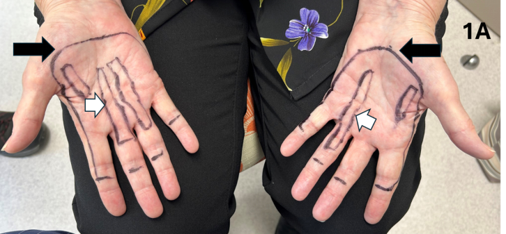
Figure 1A Clinical mark-up of both hands in preparation for VMAT planning, with the intention to treat the entire PA. The white arrows indicate the palpable nodules and cords, while the black marks represent the field borders. Note how the field borders follow the sides of the palm and, distally, extend between the PIP and DIP joints. The black arrows indicate the most medial extent of the field, corresponding to the location of the carpal tunnel. Irradiating more medially carries the risk of exacerbating the carpal tunnel syndrome.
Figure 3B: Contours at different levels in one hand.
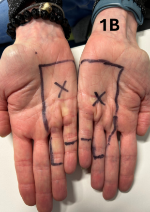
Figure 1B Treating a partial volume of the PA. This photo shows the clinical mark-up of both hands in preparation for VMAT planning, with the intention to treat only the medial parts of both the PAs. The black crosses indicated the locations where the RO has requested in vivo dosimetry (IVDs).
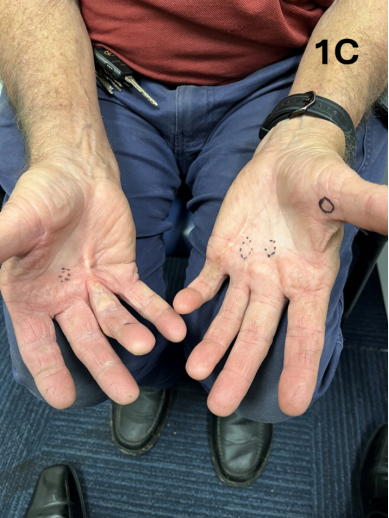
Figure 1C Nodules are present in both hands, identified by black circles. Significant fixed flexion deformity of the fourth (ring) and fifth fingers is noted. The hands were graded as at least Tubiana stage 2, and definitive RT was not considered appropriate.
Patient set-up: The field outline is delineated according to departmental practice to ensure sufficient capture on planning computer tomography (CT). This enables accurate contouring.
The patient is then positioned prone on the CT couch with a Type S-Overlay™ (CQ Medical, Orange City, Iowa, USA). One or both hands — if both are being treated— are extended superiorly above the head in what in what is referred to as the ‘Superman position’. The non-treatment hand is kept by the side of the body, outside the CT scan field. The hand(s) are then rested on a Moldcare® cushion (ALCARE, Tokyo, Japan) and secured in place by a thermoplastic S-Type™ head mask (Klarity Medical, Heath, Ohio, USA), typically used as a mask for head and neck treatment. Care is taken to ensure there is no movement of the delineation devices while positioning into the prone position. No bolus is used.
Figure 2: Detail of patient set-up at planning CT stage.
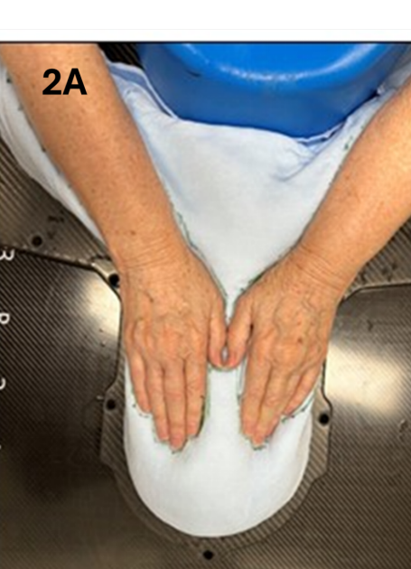
Figure 2A Immobilisation equipment used for bilateral hand treatment. The patient is positioned prone with the hands extended above the head in the Superman position. If only one hand is being treated, the untreated hand is positioned to the side of the body to keep it clear of the radiation field. The hand(s) are pressed into the Moldcare® cushion to ensure stability and reproducibility.
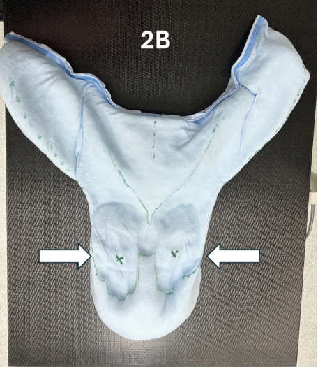
Figure 2B The Moldcare® cushion after the patient has pressed their hands into it. The white arrows indicate the green marks made by the RO on the palms, denoting the sites for IVD.
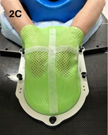
Figure 2C The hands are secured in place by a thermoplastic S-Type™ head mask, typically used for head and neck treatment. Care is taken to ensure there is no movement of the delineation devices during repositioning into the prone position. No bolus is used.
CT scan: The planning CT is then performed from the tips of fingers to the wrists using a slice thickness of 2 mm and a field of view (FOV) of 500 mm, covering the entire treatment area, including any immobilisation devices used. The scan is then exported to the treatment planning system for contouring by the RO.
Contouring by the RO: The RO contours the CT following the clinical marks shown in Figure 1A. Assuming no Garrod’s nodes are present, the distal border of the CTV lies a few millimetres above the PIP joints of each finger. The proximal border corresponds to the distal edge of the carpal tunnel. Medially, the border follows the anteromedial side of the hand, while laterally it covers most of the thenar eminence and extends distally along the anterolateral side of the index finger to a point a few millimetres above the PIP joint.
On the axial slices the CTV includes the skin and, at depth, at least contacts the tendons of the fingers, as is recommended in surgery.24 This depth varies along the volume from proximal to distal, becoming more superficial distally (Figure 3). The marked nodules can often be seen on the CT scan. An expansion to the PTV is then performed depending on departmental practice. In our department, where daily CBCTs are conducted for treatment verification, a two-millimetre margin is used.
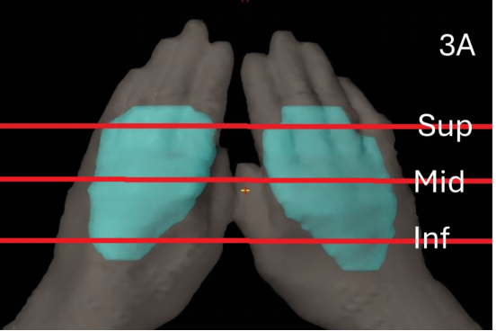
Figure 3A Scout film showing where the contours and dosimetry were taken. The PTVs are shown in blue. The superior level (Sup) was taken 1 cm inferior to the top of the PTVs; the inferior level (Inf) was taken 1 cm superior to the inferior edge of the PTVs, and the middle level (Mid) was taken from a slice midway between these two points.
Figure 3B: Contours at different levels in one hand.
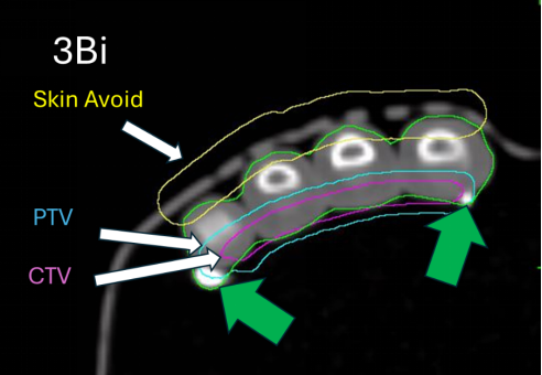
Figure 3Bi The clinical target volume (CTV) is indicated in pink, the planning target volume (PTV) in blue, and the skin avoid (SA) volume in yellow, as described in the text at 1 cm inferior to the superior level (SUP) of the PTV. The short green arrows point to white dots which are cross-sections of continuous material that is superimposed on the marks made by the RO to assist contour delineation.
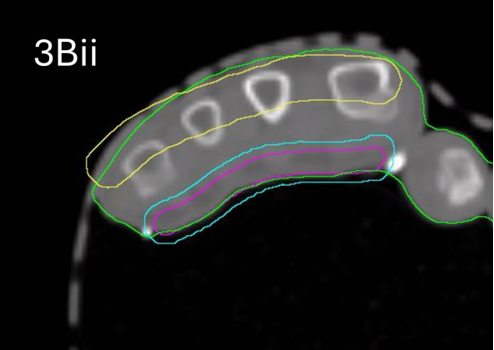
Figure 3Bii The CTV, PTV and SA volumes are shown as described in the text at the middle level (mid) of the PT.
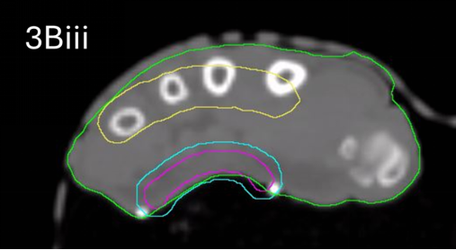
Figure 3Biii The CTV, PTV and SA volumes are shown as described at 1 cm superior to the inferior level (inf) of the PTV.
Figure 3C: The dosimetry at 95% of the prescription dose of 30 Gy.
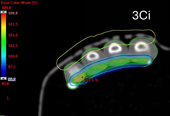
Figure 3Ci The 95% dosimetry at 1 cm inferior to the superior (sup) level of the PTV.
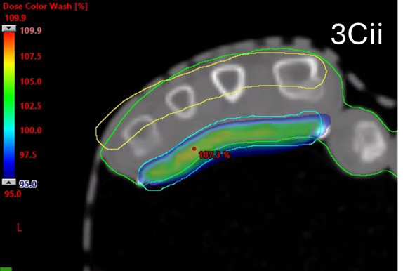
Figure 3Cii The 95% dosimetry at 1 cm at the middle (mid) level of the PTV.
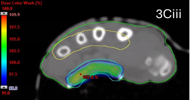
Figure 3Ciii The 95% dosimetry at 1 cm superior to the inferior (inf) level of the PTV.
Figure 3D: The dosimetry at 50% of the prescription dose of 30 Gy.

Figure 3Di The 50% dosimetry at 1 cm inferior to the superior (sup) level of the PTV.
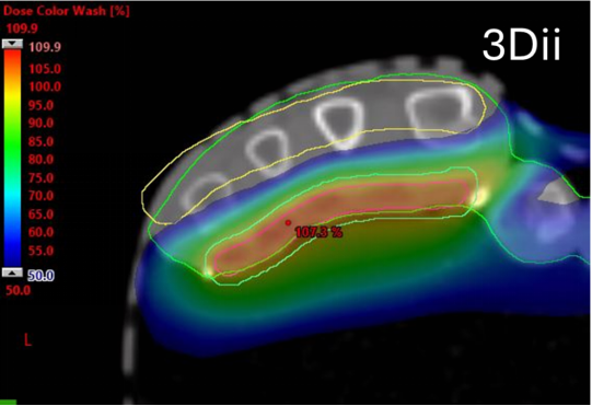
Figure 3Dii The 50% dosimetry at 1 cm at the middle (mid) level of the PTV.
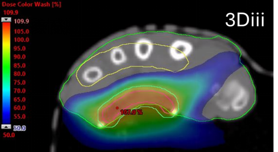
Figure 3Diii The 50% dosimetry at 1 cm superior to the inferior (inf) level of the PTV.
The RO then contours a volume called “skin avoid” (SA), which is present on every slice containing the PTV. This volume is contoured using a 10-millimetre diameter brush tool and is based on the skin of the dorsum of the hand, mirroring the palmar-facing PTV. The SA is always at least five millimetres from the PTV and may extend outside the body. This structure assists the planners to avoid irradiating the dorsum of the hand.
RT Planning: The prescribed dose to the PTV is 30 Gy in 10 fractions. The current dose constraint of the SA is a mean dose less than 8 Gy and a maximum dose of 15 Gy. This is below the threshold at which permanent alopecia is observed in healthy adults.25
Following RO contouring and prescription, a VMAT plan is generated using the Varian Eclipse Treatment Planning System, version 16.01.10, with the pre-configured Halcyon model. The dose calculation algorithm used is AcurosXB, version 16.1 (Varian Medical Systems, Palo Alto). AcurosXB is a dose calculation algorithm used in RT, specifically with the Varian Eclipse Treatment Planning System. It is designed to enhance dose calculations in heterogeneous media, such as at the soft tissue to bone interface, by more accurately modelling the interaction of radiation with different tissue types. The energy used is a 6 MV flattening filter free (FFF) photon beam with a typical depth of maximum dose (Dmax) of 1.2 centimetres (cm). Dmax refers to the depth within tissue where the radiation beam reaches its peak and delivers its highest dose before decreasing due to absorption and attenuation. The beam quality, measured as the tissue-phantom ratio at depths of 20 cm and 10 cm (TPR20/10), is 0.626. This TPR20/10 value of 0.626 indicates the relative depth dose of a photon beam in a water phantom. Specifically, it represents the ratio of the absorbed dose at a depth of 20 cm to the absorbed dose at a depth of 10 cm for a 10 x 10 cm field size at a source-to-surface distance (SSD) of 100 cm. A higher TPR20/10 value suggests a higher energy photon beam. A TPR20/10 value of 0.626 is typical of a high-energy photon beam commonly used in radiotherapy and reflects the beam’s penetrating ability.
To achieve optimal dose distribution, several optimisation structures are created in addition to the SA structure. An intermediate ring, a middle ring, and an outer ring are typically added. The purpose of these optimisation structures is to enhance dorsal skin sparing, improve dose uniformity within the target, and reduce the D5 Gy spillage. Bolus is not used even though the PTV usually includes the palmar skin. This is because the treatment area lies directly against the Moldcare® cushion, which has a density of approximately 0.15g/cm3 and a thickness of 5.5 cm—equivalent to a water-equivalent depth of 0.8 cm. This build-up depth is usually sufficient to achieve the prescribed dose at the surface of the palm, particularly as the beam is somewhat tangential to the palm’s surface. The beam’s Dmax is then more superficial.26 Bolus also increases the complexity of daily set-up and could compromise treatment reproducibility by introducing random air pockets between the bolus and the treatment area due to the intricate shape of the hand.
The dose is then calculated, the plan is accepted by the RO once appropriate, and quality assurance is carried out to ensure that the plan generated by the computer can be accurately delivered by the LA. Once these steps are complete, treatment can begin. Rings should be removed prior to treatment initiation and remain off until two weeks after the completion of each treatment phase.
RT treatment: A maximum dose rate of 800 monitor units per minute (MU/min) is utilised. Patients are treated on the departmental Varian Halcyon linear accelerator (Varian Medical Systems Inc, Palo Alto, California, USA). Prior to each treatment, a kV CBCT is acquired to ensure accurate patient positioning. Owing to the use of the Moldcare® cushion and masks, treatment setup is generally straight forward, and the need for re-setup after imaging is extremely rare. The average number of monitor units per fraction was 1600MU, with an average beam-on time of approximately 90 seconds.
Follow up: Patients are seen for on-treatment reviews (OTRs) by the RO during each week of treatment. They are also reviewed by nursing staff. The purpose of both reviews is to assess and manage skin side effects such as erythema, and to monitor for swelling, which can affect dosimetry. Swelling is relatively common in this dependent limb and usually peaks approximately one week after each week of treatment. Rarely, a sling may be required to keep the hand elevated above the heart to avoid further swelling. Other side effects are minimal and typically peak in the week following treatment completion.
The patient is reviewed at six weeks post-RT and again one year after treatment. Lingering acute side effects, along with any late effects such as anhidrosis, are documented. Patients are once again counselled about the potential for rare side effects, such as RIM, and encouraged to return if any issues arise. They are then usually discharged into the care of their primary physician or hand surgeon.
In-vivo dosimetry (IVD) was performed on the first two patients to confirm the surface dose of the treated area. The film was always placed in the centre of the PTV on the skin. Dose was measured at the first fraction. The first patient received treatment to a partial volume of one hand.
The dosimeter used was the Gafchromic™ external beam therapy (EBT3) film (Ashland, New Jersey, USA), a tissue-equivalent planar dosimeter. With a tissue-equivalent depth of 0.5mm and high spatial resolution, the EBT3 film was deemed suitable for IVD measurements without introducing significant changes to the surface dose. The uncertainty associated with the IVD measurement process using EBT3 was estimated to be ± 5.0% within the authors’ radiation oncology department. Upon completion of treatment, the film was removed and scanned on a film scanner (Epson, Suwa, Nagano, Japan), and the dose reading was determined from the pre-established calibration curve.
All patients consented to having their de-identified data used for this study. Table 1 outlines the baseline characteristics of the first 10 patients, numbered chronologically in the order their treatment was delivered. Patient 1 completed RT in December 2023, and the final patient (Patient 10) was treated in June 2025.
Table 1 shows that there were seven males and three females in the first 10 consecutive patients. The average age was 72 years (range: 63-78). Seven patients were referred by GP’s, and three by specialists. None had risk factors for diabetes, smoking, or excessive alcohol consumption. All were found to have underestimated the extent of their disease.
|
Pt No.
|
Age (yrs) / Sex/Ref |
Areas treated |
FU (Mths) |
Tub pre RT |
Tub post-RT |
Pain Pre/post-RT |
Notes |
|
1 |
78/M/GP |
R hand (Partial) |
18 |
Right 1 |
Stable |
No/No |
|
|
2 |
77/F/GP |
Bilat hands
|
18 |
Right 1 Left 0 |
Stable |
Yes/No |
Foot |
|
3 |
70/M/Spec |
L hand (Partial) |
13 |
Tub 0 |
Stable |
Yes/No |
|
|
4 |
69/M/GP |
Bilat hands (Partial) |
10 |
Right 1 Left 0 |
Right 0 Left 0 |
No/No |
|
|
5 |
73/M/GP |
Bilat hands (Partial) |
2 |
Right 0 Left 1 |
Stable |
Yes/No |
Non dom worse |
|
6 |
76/F/Spec |
Bilat hands |
7 |
Right 0 Left 1 |
Stable |
Yes/No |
Non dom worse |
|
7 |
70/M/Spec |
Bilat hands |
7 |
Right 0 Left 0 |
Stable |
Yes/No |
|
|
8 |
71/F/GP |
Bilat hands
|
7 |
Right 0 Left 0 |
Stable |
Yes/No |
Foot |
|
9 |
68/M/GP |
Bilat hands (Partial) |
2 |
Right 0 Left 0 |
Stable |
Yes/No |
|
|
10 |
63/M/GP |
Bilat hands (Partial) |
2 |
Right 0 Left 1 |
Stable |
No/No |
Non dom worse |
|
Totals/Ave |
72(63-78) 7M/3F 7GP/3Spec |
4 Bilat/4 Bilat Partial/2 Partial |
8.6 Mths |
13 x 0 and 5 x 1/18 hands |
17 hands stable; 1 improved |
7 with pain resolved |
2 feet; 3/8 Non dom worse |
Table 1 Patient and outcome characteristics
Pt, patient; No., number; yrs, years; M, male; F, female; GP, general practitioner referred; Spec, specialist referred; Bilat, Bilateral; FU, follow up; Mths, months; Tub – Tubiana; RT, radiotherapy; R, Right; L, Left; Ave, average; Dom, dominant hand; Non dom, non-dominant hand.
There were four cases of bilateral hands that were treated to the entire PA, and four that were treated to partial volumes of the PA. In two patients, only one hand was treated, both to partial volumes, giving a total of 18 treated hands. The average follow up was 8.6 months. Of the 18 hands treated, 13 were classified as Tubiana stage 0 and five as stage 1. These were considered appropriate candidates for RT.
At the sixth week follow-up post-RT, 17 hands remained stable, and one showed improvement from Tubiana stage 1 to stage 0. Of the seven patients who reported pain prior to RT, all experienced resolution of pain by six weeks. Other notable findings included two of the 10 patients also having symptomatic LD disease, which was treated concurrently. Interestingly, neither patient associated their symptomatic LD with their DD. Three of the eight patients who underwent bilateral treatment reported more severe symptoms in their non-dominant hand.
Table 2 shows the IVD results. For the first two patients treated at the authors’ oncology department, the average dose difference between the planned and the measured dose was 1.1% (standard deviation = 1.9%), well within the measurement uncertainty of ± 5.0%. This finding supports the idea that the Moldcare® cushion provides sufficient buildup for the 6MV FFF photon beam, removing the need for bolus. Based on the excellent concordance between the planned and measured doses, IVD was discontinued after the first two patients. The results justified the decision to not use bolus.
|
Pt no. and hand |
Measured Dose (Gy) |
TPS Dose (Gy) |
% Difference |
|
1 right hand |
3 |
2.98 |
0.67% |
|
2 left hand |
3.01 |
2.98 |
1.01% |
|
2 right hand |
3.05 |
3 |
1.67% |
|
Total/Ave |
3.02 |
2.99 |
1.12% |
Table 2 In-vivo dosimetry
Pt, patient; No., number; Ave, average; Gy, Gray; TPS, treatment planning system
RT planning metrics
Four patients—one male and three females— had the entire PA of both hands treated, meaning that a total of eight hands received treatment to the entire PA. The average PTV volume was 63 cubic centimetres (cc) (range: 40-85 cc). As expected, females had smaller average PTV volumes compared to males (82 cc [range: 78–85 cc] vs 59 cc [range: 40–71 cc], respectively. SA volumes for hands treated to the entire PA averaged 57 cc (range: 26-102 cc). Again, in line with anatomical expectations, males had larger SA volumes than females (92 cc [range: 82–102 cc] versus 46 cc [range: 26–66 cc]), respectively.
Four patients had partial volumes of both hands treated, and two patients had partial volumes of one hand treated, resulting in a total of 10 hands receiving partial PA treatment. The average PTV size for these cases was 46 cc (range: 27-64 cc).
The mean PTV dose was 30 Gy for all hands. The mean dose to the SA structure for bilateral hands was 12 Gy. Typically, three arcs were used, except for Patients 2 and 6 who had bilateral whole PA treatment with two arcs each. It was found that using three arcs resulted in a steeper dose gradient compared to two arcs. Given that the Halcyon has a faster gantry rotation speed than a conventional linear accelerator, increasing the number of arcs has minimal downside.
The mean monitor units (MU) for bilateral treatment to the whole PA (8 hands) was 1,145 (range: 879-1666), for bilateral partial PA (8 hands) it was 1786 (range: 1280-2182), and for two single partial hands it was 754 (range: 725-782). It was noted that the average MU for bilateral partial PA treatment was higher than that for bilaterial whole PA. This was likely due to the increased complexity of two of the bilateral partial PA plans where greater effort was made to keep the dose to the SA minimal. As a result, these two bilateral plans required significantly higher MU compared to others, which raised the overall average for the bilateral PA group. Importantly, the increase of MU and decrease in dose to the SA must be balanced against beam deliverability. As MU and plan complexity increase, the robustness of patient setup becomes increasingly critical to ensure accurate and reproducible delivery.
The dose constraints for the SA volumes were refined over time, decreasing from a mean dose of 15 Gy and maximum of 30 Gy for the first patient to 8 Gy and 15 Gy, respectively, by patient 10. This adjustment aimed to avoid the risk of any long-term lymphoedema in the treated hands. Only one patient had lingering lymphoedema at the last follow up, seven months post-treatment, and remained unable to wear her rings despite referral to a lymphoedema specialist. Her dose constraints for the SA volumes were a mean of 12 Gy and maximum of 20 Gy.
This paper describes the development and initial experience of using VMAT for the treatment of DD in the first 10 consecutive patients referred to our department. All patients referred were considered appropriate from treatment. In total, 18 hands were treated—eight to the full PA and ten to partial volumes, and RT was delivered as planned. The average follow-up was 8.6 months. Seventeen hands remained stable, and one showed clinical improvement and was downstaged from Tubiana stage 1 to stage 0. Notably, all seven patients who reported painful nodules prior to RT experienced complete resolution of pain by six weeks post-RT, representing a significant added benefit. Only one patient experienced a lingering late toxicity— persistent lymphoedema at seven months post-RT—sufficient to prevent wearing rings, despite referral to a lymphoedema specialist.
Some interesting findings emerged from this initial cohort of patients. Most were referred by general practitioners, none presented with the usual risk factors associated with DD, and all underestimated the extent of their disease, underscoring the importance of a thorough clinical assessment. Among the eight patients who had bilateral treatment, three reported worse symptoms in their non-dominant hand. Furthermore, two out of ten (20%) patients had synchronous symptomatic LD which they did not associate with their DD. Both cases of LD were treated at the same time with RT, resulting in symptom resolution. This finding highlights the importance of enquiring about foot symptoms and examining the feet of patients with DD who attend the department for RT assessment.
To our knowledge, this is the first published report to describe a technique and initial experience of treating DD with VMAT. The technique evolved iteratively, as the planning team was able to decrease the dose to the SA volume through a varied planning approach. The use of three ring structures stepped at intervals from the PTV to increase dose drop off ensured a controlled gradient. Compared with conventional radiotherapy, achieving a steep dose gradient to significantly reduce the SA dose is vital, and this was best accomplished using an aggressive normal tissue objective (priority 100, distance from target boarder 1 cm, start dose 105%, end dose 10% and fall-off 0.35).
Another planning approach is the use of flexible priorities during optimisation to balance the PTV coverage and dose gradient. As the multi-resolution (MR) levels progress, the priorities can be adjusted to achieve an optimal plan. Specifically, during the early MR levels (MR 1-2), higher objective priorities were assigned to the ring structures and to the SA to bring down the SA dose. As the MR level progressed to MR 3-4, the PTV coverage priority was increased by a factor of two to ensure adequate coverage without compromising the low SA dose.
A further practical advantage identified during our experience was that the use of bolus was unnecessary. IVD showed that the Moldcare® cushion provided sufficient dose build-up. This finding meant a more stable treatment position and faster set-up time.
With this technique, treating only a partial volume of the PA carries the risk of field- edge recurrence and the development of new symptomatic nodules just outside the RT volume.27,28 Planning must therefore anticipate the potential need for future field matching, should re-treatment be required. Patients should be advised to avoid activities that may increase microtrauma to the hand, which could stimulate disease progression.
The decision to treat the entire PA versus a partial volume to cover symptomatic nodules can be likened to the spectrum of surgical approaches, which vary depending on disease severity and therapeutic intent. Surgical techniques have been well summarised by Ruettermann et al. and escalate from steroid injections into the hard lump to soften, flatten and relieve pain from the nodules to collagenase injections, percutaneous needle fasciotomy, limited fasciectomy and ultimately, extensive fasciectomy. The trade off with surgery is that as the invasiveness of the procedure increases, the recurrence rate decreases but the complication rate rises. The reported overall recurrence rate at five years varies widely, from 2% to 73%.29
Other RT modalities have been employed in the treatment of DD. Yunes, using 6 MeV, aimed to place the 90% isodose line at the base of the PA in the palm and at the level of the periosteum of the fingers more distally. This depth modulation with electrons was achieved using a custom three-dimensional bolus material that was produced in-house.30 Seegenschmiedt et al.,9 in an excellent randomised trial, demonstrated that orthovoltage RT could be just as effective, supporting a range of RT options for DD depending on equipment availability and institutional expertise.
This study has several limitations. It is a single institution, retrospective analysis of a small patient cohort with short follow-up. Further research with larger studies is warranted. Nonetheless, there is merit in presenting this VMAT-based technique for centres without access to other RT modalities, such as electron or orthovoltage therapy, where VMAT may be the only available option.
This article describes a technique for successfully treating Dupuytren’s disease using volumetric modulated arc therapy. In total, 18 hands were treated. Of these, 13 were Tubiana stage 0 and five were Tubiana stage 1, indicating appropriate selection for definitive radiotherapy.
Several notable observations emerged. None of the patients had typical risk factors and all underestimated the extent of their disease. Encouragingly, all seven patients who reported pain prior to radiotherapy experienced complete resolution by six weeks post-treatment. Two patients were diagnosed with symptomatic Ledderhose disease at the time of their Dupuytren’s referral. These two patients were treated simultaneously for both conditions and did not recognise the connection between them. Interestingly, in three of the eight patients receiving bilateral treatment, symptoms were more severe in their non-dominant hand. In-vivo dosimetry confirmed that bolus was not necessary when using a Moldcare® cushion.
This retrospective, single institution study is limited by small patient numbers and short follow up. However, the findings may be of value to radiotherapy departments that lack traditional radiotherapy modalities. Further study is warranted.
The authors wish to thank their colleagues for referring the patients who participated in this series, as well as the patients themselves. We also thank Icon Cancer Centre for funding the open access publication of this manuscript. Finally, we would like to thank Aileen Eiszele of A&L Medical Communications for editing and preparing this manuscript for submission.
The authors have no conflicts of interest to declare

©2025 Fogarty, et al. This is an open access article distributed under the terms of the, which permits unrestricted use, distribution, and build upon your work non-commercially.