International Journal of
eISSN: 2574-8084


Educational Review Volume 9 Issue 2
1ICON Cancer Centre, Revesby, New South Wales 2122, Australia
2Strategic Investment and Clinical Care, ICON Cancer Centre, Queensland 4101, Australia
Correspondence: Gerald B. Fogarty, ICON Cancer Centre, Revesby, New South Wales 2122, Australia
Received: June 27, 2022 | Published: July 11, 2022
Citation: Fogarty GB, Sutton A, Ni R, et al. A scientific approach to skin radiotherapy nursing Article 1 - The role of the nurse in patient assessment. Int J Radiol Radiat Ther. 2022;9(2):48-54. DOI: 10.15406/ijrrt.2022.09.00324
Nurses play an important role in the care of patients treated with radiotherapy (RT) for skin conditions, such as skin cancers. A series of four articles will aim to supplement general oncology nurse training by providing a scientific basis to better understand the skin and its diseases, the use of RT in skin, and the management of necessary acute skin RT reactions to achieve best patient outcomes.
This first article focuses on the role of the nurse within the radiation oncology department and the initial patient assessment. It enables the nurse to develop an appreciation of the unique nature of patients undergoing RT to the skin and provides recommendations on nursing assessment, patient examination, and optimal care of the skin RT patient. It also highlights the psychological impact of a skin cancer diagnosis on the patient.
The second article in this series of four revises the anatomy, physiology, pathologies and radiobiology of skin. The third describes the skin RT prescription and plan, especially in relation to volume and dose; and the fourth shows how the nurse, by applying the knowledge from the previous articles, can predict, explain and care for acute side effects that may arise during a course of skin radiotherapy.
Keywords: radiotherapy, radiobiology, superficial radiotherapy, volumetric modulated arc therapy, skin, skin cancer, nursing, review
High quality nursing is essential to achieve the best outcomes from skin radiotherapy (RT). Oncology nurses spend a significant amount of time, perhaps the most of any RT craft group, with skin RT patients, especially when applying dressings and imparting education. In Australia, subspeciality nurse training in RT is achieved on the job at each institution and through agencies such as the Cancer Institute of New South Wales EviQ resource.1 EviQ is a free internet resource of evidence-based, consensus-driven cancer treatment protocols and information developed for the Australian context. For nurses, EviQ has developed the Radiation Oncology Nursing Knowledge and Skills Framework2 to support a standardised level of knowledge and clinical skill for the safe care of patients receiving RT.
To supplement general RT training and education, we provide here a series of four inter-related and practical review articles to support nurses to achieve clinical excellence in the care of patients undergoing RT for skin conditions.
This first article examines the essential role of the nurse within the RT department. It then focuses on the nurse’s role in the management of the skin RT patient during treatment and offers patient assessment recommendations to ensure optimal treatment completion. Article two revises the anatomy and physiology of skin – its structure and how it functions. It then highlights key skin pathologies and how the radiobiology of skin and its diseases impacts the skin’s response to RT. The third article describes the skin RT prescription and plan, especially in relation to volume and dose. The fourth paper shows how the nurse, by applying the knowledge from the previous articles, can predict, explain and care for any acute side effects that may arise (Tables 1 & 2).
Article |
Topic |
1 |
The role of the nurse in patient assessment and the RT department. |
2 |
Anatomy, physiology, pathologies and radiobiologies of skin and its diseases. |
3 |
The skin RT prescription and plan – focus on volume and dose. |
4 |
Knowledge application – using the information gained from these articles to predict, explain and care for acute skin side effects in skin RT patients. |
Table 1 Scope of the four articles
Concept |
Explanation |
Therapeutic ratio |
A treatment modality with a better therapeutic ratio has increased cure of cancer and less side effects in normal tissues compared with another with a worse therapeutic ratio. |
Consequential RT late effects |
Permanent side effects that may develop usually because of substandard care of acute RT effects. |
Toxicity prediction |
Predicting RT toxicity from the RT prescription and RT plan. |
Nurse role |
The role of the nurse in the skin RT patient journey. |
VMAT |
Volumetric modulated arc therapy - the latest in generally available megavoltage treatment. |
ESFC |
Extensive skin field cancerisation - actinic change measuring over 50cm2 (about the size of an adult’s palm). |
Quality of life |
The standard of health, comfort, and happiness experienced by an individual or group. |
Table 2 Concepts used in this article
Radiotherapy has been used for the successful treatment of skin diseases for over 100 years.3 The incidence of skin cancer is increasing,4 especially in the aged.5 The aged can have mobility issues, nutritional deficits and comorbidities that need more nursing care. The use of RT is increasing in this population for multiple reasons, some of which are described here. Compared with surgery, RT does not need an anaesthetic, reversal of anticoagulation, or weeks in bed to allow a graft to take on a dependent limb. Compared with topical therapies, such as creams, a course of RT is relatively short and has less compliance issues; that is, there is less risk that treatment is directed to the wrong area or not administered at all. Patients may prefer RT as, unlike surgery, there is no sacrifice of normal tissue and therefore a better functional and cosmetic outcome (Figure 1). Table 3 provides a comparison of RT, surgery and topical therapy for skin lesions.
Factor |
Item |
RT |
RT |
Sx |
Sx |
Top |
Top |
Patient |
|
Adv |
Dis |
Adv |
Dis |
Adv |
Dis |
|
Tissue loss |
No |
|
|
Yes |
No |
|
|
Comorbidity impact |
Minor |
|
|
Major |
Minor |
|
|
|
|
|
|
|
|
|
Tumour |
|
|
|
|
|
|
|
|
Frequency of application |
|
Yes |
No |
|
|
Yes |
|
Histology report |
|
No |
Yes |
|
|
No |
|
|
|
|
|
|
|
|
Treatment |
|
|
|
|
|
|
|
|
Anaesthetic |
No |
|
|
Yes |
No |
|
|
Reverse coag |
No |
|
|
Yes |
No |
|
|
Bed rest for graft to take* |
No |
|
|
Yes |
No |
|
|
Self-administration |
No |
|
No |
|
|
Yes |
Table 3 Comparison of RT, surgery and topical therapy for skin lesions
*legs
Abbreviations: RT, Radiotherapy; Sx, Surgery; Top, Topical therapy; Freq, Frequency; Adv, Advantages
Dis, Disadvantages; Coag, Coagulation
RT has improved. There has been an increase in the understanding of the radiobiology of skin and malignant6,7,8 and benign9,10 diseases. Modern techniques, such as volumetric modulated arc therapy (VMAT),11,12 enable RT delivery with more conformity and homogeneity of radiation dose to the target, thereby increasing what is known as the therapeutic ratio. A better therapeutic ratio describes a (better) treatment modality offering increased cure with less side effects in normal tissues compared with another (which has a worse therapeutic ratio).
Nurses play a vital role in the delivery of RT for skin disease. Specifically, they initially assess and educate the patient, usually at the RT planning stage. Nurses can allay concerns and ensure patient safety and comfort as treatment commences. Nurses then follow the patient’s progress and monitor the patient closely for side effects throughout the fractionated course of therapy. Following treatment completion, they then attend to the successful resolution of skin acute effects, which may take weeks and even months to resolve. Radiation nurses often share the latter load by referring the patient to their community nursing colleagues. This enhances patient convenience once treatment is finished and helps to ensure that no consequential RT late effects develop following RT.13 Consequential RT late effects are permanent side effects that can develop, usually due to substandard care of acute RT effects.
The general role of the radiotherapy nurse
Nurses act a patient advocate in RT, as they do in all workplace environments. They spend a significant amount of time, perhaps the most of any RT interdisciplinary team, with skin RT patients, especially when applying dressings and imparting education. Nurses escalate issues and concerns to radiation therapists, physicists, and radiation oncologists (ROs) when needed. These RT craft groups have specific training in RT and sometimes years of experience in dealing with skin, which may not necessarily be the case for some nurses. Nurses, through no fault of their own, may therefore not be sufficiently versed in the science behind the effects of RT on the skin and the protocols they are asked to follow. This may lead to nurses being unable to explain to the inquisitive patient, or non-RT hospital staff, the scientific basis underpinning the protocols, resulting in a lack of trust and compliance. More scientific knowledge may empower nurses to identify when and why unexpected RT reactions in skin are occurring so they can solve these themselves or escalate appropriately.
Nurses are good at creating and following skin care protocols. From their training, nurses bring a pragmatic yet personalised and patient-centric approach to protocol application. Two patients may be having the same treatment for the same disease, but their individual characteristics, such as mobility, ability to self-care and cognitive function etc., may differ. Nurses can take individual needs into account and finesse protocols so that they can be applied appropriately but differently. The care result also depends on the ability of the nurse to feel confident and supported in their care provision by the rest of the RT team.
Nurses play a significant role in the RT department. RT delivery involves many people, and its safe and effective delivery is dependent on interdisciplinary collaboration, trust, and efficient and effective communication. Nurses bring a holistic approach to care that often transpires into relationships with other staff. They can provide the oil that lubricates the communication between the other RT craft groups. They make an important contribution to the welfare of RT staff and workplace culture, and therefore can impact the productivity of the RT department.
Nurses also play a significant role between the RT department and the rest of the hospital. Nurses can identify with other nurses throughout the hospital and therefore provide an important connection14 between departments. They help ensure the acceptance of the RT department within the whole hospital, especially when it comes to effective communication. This is particularly important when inpatients are receiving RT.
Knowing the patient receiving RT to the skin
The patient receiving RT to the skin may differ from the usual RT patient in several ways. Patients presenting for skin RT vary greatly. For example, they can range from a person in their 20’s presenting post-operatively for immediate RT (PORT) following excision of a persistent benign keloid where the first fraction is best given within 24 hours of surgery to ensure success; through to the centarian with severely sun damaged skin receiving palliative RT for symptoms from an inoperable keratinocyte cancer. Skin conditions can affect virtually every body site, and the impact of irradiating skin at one site (e.g., the nose) is different from another (e.g., the back).
The nursing assessment: Nurses have a refreshing holistic approach to patients. However, the radiation oncology nurse may only first meet the patient for consultation during the planning visit. This usually occurs after the treatment course has been explained and the patient has consented to treatment. The radiation oncologist may dictate a letter explaining the reason for therapy back to the referring doctor or for the patient’s notes, but the letter may not be available at the time of the nurse’s initial consultation with the patient.
The nursing assessment requires the patient’s history to be documented and an examination. If a ‘health history’ document is not available, the nurse may need to piece together the reasons for treatment from the referral letter and a conversation with the patient. The patient, often elderly with comorbidities severe enough to preclude surgery, may also not be the best historian regarding their own health. This situation can be challenging and make it appear to the patient’s family or carer that the treatment team does not communicate amongst each other given that the patient’s history and treatment scenario does not seem to have been relayed from the doctor to the nurse.
Important items to document in the patient’s history include nutrition and who provides it, transport and who organises it, and planned accommodation for out of area or rural patients. These are essential general factors that can impact the RT prescription and treatment success. The nurse should enquire about any other treatments the patient may be using such as fluorouracil cream, often prescribed by dermatologists in this patient cohort, to ensure that radio-sensitising agents are not inadvertently used concurrently. Smoking and inadequately controlled diabetes may impact wound healing and are best understood prior to RT so that they can be considered. Referrals to other specialists may also need to be generated. Patients should be asked if they or their family have a history of cancers, especially skin cancers, and what previous treatments were administered. The level of help available to the patient at home and the patient’s ability to self-care may determine if more care oversight is needed. Previous or familial increased toxicity to RT is rare but worth discussing. A positive answer may indicate a problem with deoxyribose nucleic acid (DNA) fragility or repair.
Patient examination should include baseline observations, especially weight, as weight loss during treatment may cause earlier than expected acute toxicity and necessitate a break or early cessation, risking RT failure. Skin thinness and fragility should prompt more regular follow-up on treatment. It is also important to ensure that the patient can see and attend to the area being treated, or that there is someone who can assist; for example, with a lesion on the back. As with all RT, knowing if the patient has a pacemaker and how close it is to the RT treatment volume is vital as RT can interfere with the function of some of these devices. As per best practice guidelines, a pacemaker check should be carried out prior to RT to capture baseline functionality depending on departmental policy.
Skin lends itself to observation and the main imaging modality in skin is photography. Photos are normally taken by the RT team and perhaps by the radiation oncologist. Their function, however, is often with planning, treatment set up and clinical needs in mind rather than those of regular nursing assessment. The technique for taking adequate photos with clear reproducible measurements to aid the nursing treatment assessment during treatment is beyond the scope of this work but it may need to be considered and resourced at a departmental level. Many cancer care organisations have clinical photography policies and procedures. Nurses need to use a consistent and recognised grading tool for assessing radiation induced skin reactions (RISRs) such as CTCAE.15 This provides consistent language and grading amongst the interdisciplinary team and is in keeping with communication safety requirements in healthcare. This is an evidence-based approach endorsed by agencies such as the Cancer Institute of NSW EviQ resource.1 Clinical photography compliments and records the clinical assessment of toxicities.
The ability to take quality photos and measurements, and to record these accurately so that they are comparable week by week, is an important nursing skill. Skin lends itself to measurement with rulers and tape measures with accurate notation. Serial photography of acute changes can help to monitor the development of reactions, especially in those who are expected to have problems. It also facilitates remote specialist skin assessment when the radiation oncologist is not present during treatment, or when the patient has completed treatment but cannot return to the treatment centre for review for a variety of reasons. Nurses need to be able to teach isolated patients and carers, who are unable to come in for regular treatment reviews following RT, to take their own photos for videoconference review.
Nursing assessment may also include blood tests. Adequate haemoglobin is needed to ensure oxygenated tissue as RT efficacy is linked to oxygenation. Poor nutrition is associated with poor wound healing. Vitamins B12, C, D and E; serum folate, calcium, magnesium, phosphate, selenium and zinc are important for healing. Deficiency can be common, especially in groups that may be at risk. At-risk groups may include people with a lower socioeconomic status, the socially isolated, or those with mobility issues. More research is needed. A HbA1c blood glucose test may help to understand diabetic severity and recent control, and a serum cotinine level can add objectivity to a history of smoking. The use of a validated tool for malnutrition screening, such as the Malnutrition Screening Tool16, and the establishment of referral pathways for patients to a dietitian when needed, are other important skills to master for the skin RT patient.
Unique nursing assessment factors for the skin patient
Psychological impact of skin cancer: Humans relate with the outside world through their skin. Skin is often exposed to the sun. UV light from the sun can cause skin cancer in susceptible individuals. Patients with skin cancer must deal with not only the impact of having cancer but also the impact of treatment-related side effects which can have functional and cosmetic effects. Sometimes these considerations can lead a patient to consider RT rather than surgery. As RT does not remove tissue, there is less impact on functional and cosmetic outcomes (Figure 1). With RT, there is no need for anaesthesia or a prolonged hospital admission during which the patient remains immobile so that a skin graft on the lower legs, for example, can take. There is also no need to alter anticoagulants prior to therapy. Some patients seek RT as they have ‘surgical fatigue,’ and are tired of painful local procedures or general anaesthesia, and of losing more tissue. Elderly patients can also shy away from general anaesthetics for surgical procedures due to a fear of cognitive side effects.
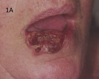
Figure 1A Large neglected cutaneous squamous cell carcinoma (cSCC) of the right lower lip. Surgical removal would require tissue replacement with the risk of neurovascular compromise.
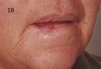
Figure 1B Six months following definitive RT. This lady retains her lip and natural lines. There is some tissue loss, but this is probably due to either over vigorous brachytherapy, a type of radiotherapy, or central necrosis which was already present when this tumour was treated.
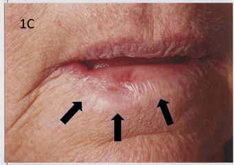
Figure 1C Close up view. Arrows indicate the line above which cancer was present. This volume of normal tissue was inside or invaded by the cancer. At the very least, this volume of tissue would have been sacrificed with surgery and may have needed replacement. Normally more tissue would be taken to ensure negative margins. Tissue replacement may have issues with adequate neuro-vascular take and movement, impacting oral continence and competence.
Figure 1 RT does not remove tissue and so has decreased impact on functional and cosmetic outcomes.
Multiplicity of lesions: Patients receiving RT for skin pathologies may have multiple lesions requiring treatment, unlike other RT patients who are dealing with only one focus, for example, definitive treatment of cancer of the prostate. These skin lesions may be synchronous and can be treated with one RT course delivering treatment to multiple sites; or they may be metachronous, appearing over time and involving multiple treatment courses. Treating multiple sites in the same course will add to lethargy and may impact self-care compliance. This may necessitate a more rigorous schedule of reviews while the patient is on treatment. The decision may be made to treat a crop of lesions in one course, provide the patient with a treatment break, and then bring the patient back to treat the remaining pre-existing lesions. This latter group of patients can become regular customers.
Some patients even have large areas of actinic field change, usually in sun exposed sites, that continually give rise to new skin cancers. These patients can be called keratinopaths. They need to have ongoing treatment to new cancers in the same area. They suffer the embarrassment of appearing with visible skin changes in public, and this may predispose them to mental health problems.17
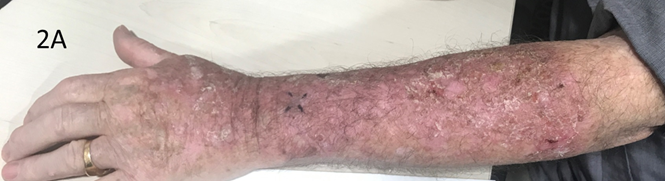
Figure 2A Some patients suffer with large areas of actinic field change of in situ or non-invasive disease usually from long term sun exposure. When over 50cm2, roughly the size of an adult’s palm, these changes can be called extensive skin field cancerisation (ESFC). These areas keep on producing new invasive cancers that need individual attention. A patient’s quality of life can be poor due to the skin being itchy and flaking.
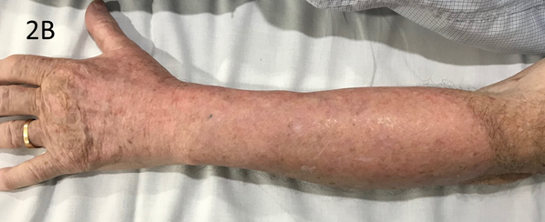
Figure 2B Six months after VMAT. There have been no new in-field lesions in this patient for over three years following VMAT.
Figure 2 A new type of RT called volumetric modulated arc therapy (VMAT) can be used for extensive skin field cancerisation (ESFC).
Regular visitors: Keratinopaths, in particular those who are immunosuppressed, may come in for so many courses of treatment that they may become regular visitors to the department. Staff may form stronger relationships with these patients and become quite attached. Some patients may eventually develop regional and metastatic disease and succumb, which can have ramifications for staff well-being.
Dose de-escalation: There is indirect evidence that skin cancer is more radiosensitive and needs less dose.18 ROs interested in dose de-escalation may try to make the RT journey for patients easier by decreasing the total dose and therefore the number of fractions, or by trying novel fractionation schemes like the adaptive split course.19,20 This is a useful approach for the frail and elderly with transport or cognitive concerns as shorter treatment is easier to facilitate and tolerate for this cohort. The rationale for doing so needs to be well communicated amongst the RT team on the understanding that this de-escalation approach can fail. More research is needed.
Endpoints of local control and functional and cosmetic outcome with less emphasis on survival: Small keratinocyte cancers will rarely kill, but if treated inappropriately can cause morbidity and poor functional and cosmetic outcomes. Some patients may select RT above other modalities such as surgery for cosmetic and functional reasons. Expectations may be high and should be discussed.
Treatment for benign disease and in situ disease: The indications for RT in benign disease such as keloids,21 rosacea,9 psoriasis,10 hidradenitis suppurativa;22 and in-situ disease, such as extramammary Paget’s disease23 and extensive skin field cancerisation,24 are growing. These patients may have different expectations, especially about survival, from those receiving RT for non-skin invasive cancers like breast and prostate cancers. Patients with benign disease may feel challenged by having to visit a cancer centre for treatment. Sharing the same waiting room with patients who are fighting to survive may be uncomfortable. Some skin patients may not seem to be aware of the suffering surrounding them in the cancer centre.
The overrepresentation of the immunosuppressed: The immunosuppressed have a worse prognosis for all cancers, but this is especially true of the cohort presenting for RT for skin cancer. They may have chronic lymphocytic leukaemia,25 human immunodeficiency virus,26 be a transplant recipient,27 or have been on immunosuppressive drugs like steroids28 for other conditions, such as some forms of arthritis, for years. Immunosuppressed patients may be overrepresented in Australia due to greater skin sun exposure given this country’s outdoor lifestyle, a high UV index, and a significant part of the population with genes from northern Europe. These patients are also prone to infection. Expert nursing care to manage broken skin due to RT is essential to avoid opportunistic infections.
The overrepresentation of the elderly: Elderly patients are overrepresented in the skin RT practice. Immunosenescence,29 or the deterioration of the immune system with age, leads to an exponential increase in skin cancers in the elderly, especially in those whose skin was sun damaged in their youth. This overrepresentation may also be because surgery is not an option in patients with advanced age due to the increased presence of co-morbidities. Skin healing is dependent on nutrition, and the elderly may have subclinical malnutrition,30 especially those that are socially isolated. Nurses must address the nourishment of these patients during RT and regularly assess their weight. Transport for the elderly is usually provided by the family who may have competing obligations for time. This can become a major issue when fractionated courses of RT are needed and may be the reason underlying the choice of a particular fractionation pattern.
Skin as the RT target organ: In skin RT, the skin is the obvious key target. This differs from other situations where the skin may be affected as a consequence of treatment, for example, RT to the breast. In skin RT, a brisk radiation induced skin reaction is to be anticipated. Managing the patient and family expectations around this is important, and a consistent approach to education based on mutual understanding between the radiation oncologist, radiation therapists and nurses is the key to successful patient management.
Nursing care continues after the completion of RT: At the end of treatment, the side effects in normal skin in the irradiated area or volume will be approaching their peak. Nursing care extends beyond the last treatment date to ensure that skin healing follows its predictable trajectory toward resolve without infection to avoid long-term fibrosis from healing by secondary intention. Patients and carers need to know that they will be followed by the RT nursing team until healing post RT is complete. Sometimes follow up care may occur in conjunction with external or community nursing services as individually required. This is different to the typical follow up care that other cancer patients undergoing RT receive. The latter may only see their radiation oncologist for review four to six weeks after the completion of treatment. With these patients, contact with nurses post RT is generally only initiated if there are concerns about a patient’s ability to self-care, if a skin reaction requiring dressings is present or anticipated, or if follow up is requested by the patient or carer. Radiation oncology treatment centres that treat skin patients need to factor skilled nursing care following the completion of RT into their budgets and schedules.
Factors in the patient’s nursing history that can predict for exaggerated acute RT side effects
Patient skin type: Some single nucleotide polymorphism (SNPs) of the MC1R gene have been shown to have more sensitivity to RT acute effects. These alleles also code a ‘red hair colour’ phenotype, like Fitzpatrick type 1 skin.31 This skin phenotype is characterised by red hair, blue eyes, white skin, freckles, a tendency to sunburn and not tan,32 and is often found in people of Celtic descent. This provides indirect evidence that in these patients the acute effects of RT on normal skin and on skin cancers may be enhanced, but more research is needed. Nurses need to be alert to the potential for earlier acute changes in these patients.
Comorbidities and Concomitant drugs: Whether collagen vascular diseases (CVDs) lead to increased acute effects, especially in the setting of hypofractionation, is controversial. Annotation of the duration of CVD, whether the disease is still active, and a history of CVD drug therapy is important. Some patients may need to be on radiation sensitising drugs during RT for a concomitant illness, for example, methotrexate for rheumatoid arthritis, hydroxyurea for certain types of chronic myelogenous leukaemia, or statins for cholesterol etc. Acute effects may occur earlier. Concurrent cytotoxic chemotherapy (CT) can cause increased side effects, but it is not used routinely in cutaneous squamous cell carcinoma (cSCC) following a successful Australian based randomised trial (RCT).32
Re-Irradiation: Acute effects can be heightened in patients who have the same area re-treated. Those who have re-irradiation soon after a previous course will be more at risk. There is some evidence that radiation effects in normal tissue are ‘forgotten’ over time.34 This may mean that reirradiation is possible in some scenarios.
Genetic predisposition: Rarely, some patients have a genetic predisposition that enhances radiation acute reactions.35,36 Patients with a history of truncated RT treatments in the past, relatives with severe acute RT reactions, or a strong family history of cancer, which may indicate problems with DNA fragility and/or repair, need to be watched closely on treatment and beyond.
Nurses play an important role in the RT of skin and its diseases. This article, the first of four, attempts to describe the important role of the nurse in the skin patient’s therapeutic journey. It also offers suggestions as to what nurses should look for during the initial nursing assessment and provides an appreciation of the unique nature of skin RT patients.
The use of RT to treat skin and its diseases is increasing due to an increased incidence of skin cancer, especially amongst an ageing population for whom surgery or topical therapy may not be appropriate or declined. In addition to the conservation of normal tissue, RT does not need an anaesthetic, reversal of anticoagulation, or weeks in bed to allow a graft to take on a dependent limb. The application of RT in skin has improved due to an increase in the understanding of radiobiology and the integration of new modern techniques that enable RT to be delivered with more conformity and homogeneity.
Nurses play a vital role in the delivery of RT. They act as patient advocates, spend significant time with patients, and help to develop and enact skin care protocols to achieve excellent outcomes, but they may suffer from a lack of specific radiation training in skin. Nurses also perform a significant communications role within and beyond the RT department.
Knowledge of the patient receiving RT to the skin is key. The initial nursing assessment of the patient requires a clinical examination and documented history.
Observation, measurement, and clinical photography are important parts of the initial assessment. Nurses should be aware of the unique factors to be considered when assessing skin RT patients. These include an understanding of the psychological impact of skin cancer and the presence of multiple lesions, or large areas of actinic field change, that continually give rise to new cancers. These keratinopaths can become regular customers.
Skin patients can have different expectations compared to other cancer patients with regards to the treatment endpoints of local control or functional and cosmetic outcomes, as opposed to survival. Those with benign disease may be challenged by having to visit a cancer centre for treatment. The immunosuppressed and elderly, with all their extra nursing complexity, are overrepresented in this cohort.
Unlike other cancers, the skin itself is the RT target. Nursing care continues beyond the last fraction of treatment. Factors that can predict an increase in acute effects include the patient’s skin type, comorbidities, concomitant medications, re-irradiation to the original site, and rarely some genetic predispositions.
The second article in this four-part series will revise the skin’s anatomy (structure) and how it functions (physiology). It will then examine specific diseases or pathologies of the skin and their ability to respond to ionising radiation (radiobiology).
The authors wish to thank Aileen Eiszele of A&L Medical Commmunications for writing assistance and for overseeing the journal submission and acceptance process. We are also grateful to Xstrahl for supporting the medical writer and open access publication.
All authors declare that there is no conflicts of interest.

©2022 Fogarty, et al. This is an open access article distributed under the terms of the, which permits unrestricted use, distribution, and build upon your work non-commercially.