International Journal of
eISSN: 2574-9862


Review Article Volume 7 Issue 1
1Department of Animal Nutrition and Husbandry, University of Veterinary Medicine and Pharmacy, Košice, Slovakia
2Department of Biology and Physiology, University of Veterinary Medicine and Pharmacy, Košice, Slovakia
3Department of Food Hygiene Technology and Safety, University of Veterinary Medicine and Pharmacy, Košice, Slovakia
4Department of Husbandry and Development of Animal Wealth, Faculty of Veterinary Medicine, Menoufia University, Menoufia, Egypt
5Department of Pathobiochemistry, Faculty of Pharmacy, Meijo University Yagotoyama, Japan
Correspondence: František Zigo, University of Veterinary Medicine and Pharmacy, Department of Animal Nutrition and Husbandry, Košice, Komenského, Slovakia, Tel +421- 908-689-722
Received: February 20, 2023 | Published: March 3, 2023
Citation: Zigo F, Ondrašovičová S, Farkašová Z, et al. Correct interpretation of carrier pigeon diseases – Part 1: Ornithosis complex. Int J Avian & Wildlife Biol. 2023;7(1):15-19. DOI: 10.15406/ijawb.2023.07.00185
The most fundamental and important topics for carrier pigeon fanciers are health and form of their competitors. It does not matter how good a pigeon is or how well fed it is, an unhealthy pigeon will never be able to win a race. One of the most common diseases of carrier pigeons during the racing season is a respiratory illness named "Ornithosis Complex". Also known as "Ornithose", "Coryza" and "One-eyed Colds" it is caused most often by a combination of Chlamydia psittaci, Pasteurella, Mycoplasma, Herpes virus and many other common bacteria such as Yersinia, Enterobacter, Streptococci and Staphylococci. Once both old and young birds have started their training and racing program, they are likely to develop an ornithosis complex, showing symptoms of swollen or puffed feathers around the ears, dry plumage, blue flesh and a disruption to moulting. Pigeons that arrive home showing signs of extreme fatigue and typical ornithosis symptoms should be treated directly with a combination of prescribed treatments, isolated from healthy birds and should not be flown to avoid spreading the disease. The study points to the fact that ornithosis complex is not a difficult problem when carrier pigeons do not overflow. It will become a big problem only after the start of the racing season, which will take away the success of many breeders.
Keywords: coryza, one-eyed colds, respiratory, symptoms, Chlamydia psittaci
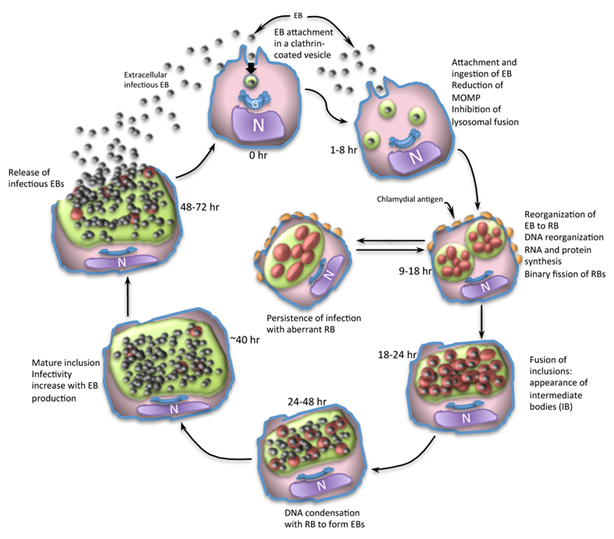
Due to the possibility that feces and nasal secretions can contain significant amounts of organisms, respiratory infections are particularly crucial in the transmission of the disease. Ornithosis complex caused most often by a combination of Chlamydia psittaci, Pasteurella, mycoplasma or viral agents. Other bacteria as E. coli, Yersinia, Enterobacter, Streptococci or Staphylococci may also play a role.1 Among the most common agents involved in this systemic disease is Chlamydia psittaci, which behaves like a virus, even though it is a bacterium, because it does not have a cell wall and lives inside the infected cells of pigeons and other birds. Ornithosis or psittacosis is the traditional names for this Gram-negative, obligate intracellular bacterium. It is a infectious disease that can affect both domestic and wild birds and is spread to people. Both horizontal and vertical pathways can spread bacteria. The horizontal transmission is the most common way of infection with three distinct forms during its life cycle: elementary body (EB), reticular body (RB) and intermediate body (IB) (Figure 1).2

Figure 1 Chlamydia life cycle.
Source: Morais et al.2
The EBs that infect the target epithelial cells connect to their surface to start the infection process. To enclose the EBs, these cells encourage the creation of a pseudopod. This bacterium prevents the vesicle from joining the cell lysosomes inside the cytoplasm. Together with the nascent inclusion, reticulate bodies replace extracellular bodies (EBs) (RB). Through binary fission, RBs replicate late in the cycle, creating both RBs and intermediate bodies (IB). Antigenic proteins are now visible on the cell surface. At this point, a chlamydia cycle arrest could start a persistent infection or continue the cycle, resulting in the formation of an elongated, abnormal RB. Before DNA condensation and RB transformation into a newly formed EB, the numerous intracytoplasmic inclusions containing bacteria within can also merge at this phase, causing the agent to evolve into intermediate bodies (IB). During EB development, the mature inclusion grows in size until it becomes infectious and is released into the extracellular area to start a new intracellular cycle. Elementary bodies, reticulate bodies, and intermediate bodies in Figure 1 are represented by the letters N, G, and IB respectively.2
The agent is excreted on faeces and ingested from the food or inhaled via aerosols. At the lungs of newly infected animals, the organism gets an infecting status becoming capable to replicate and causing clinical signs of disease. Chlamydia can be transmitted in pigeons of any age, which can be manifested not only by respiratory but also by other systemic diseases, and without proper treatment, birds may die. In some pigeons, the functionality of the gonads may be impaired leading to a decrease in the fertility of both pigeons and doves. Doves with a chlamydial ovarian infection often ovulate late or irregularly, or do not ovulate at all. If an egg is formed, chlamydia can sometimes be transferred to the egg, which can either kill the developing embryo or lead to the hatching of a weakened chick.3
Studies on Chlamydophila’s prevalence increased and psittacosis was classified as an endemic disease in Belgium and other European countries, such as France and Germany.4,5 The most concerning cases involve psittacines and pigeons, whose prevalence ranges from 16% to 81% and whose mortality is frequently exceeding 50%.5,6 The seropositivity of wild pigeons vary widely, from 12.5% to 95.6%. When this species lives in close proximity to humans in both urban and rural settings around the world, the seropositivity is concerning. Between 35.7% and 60% of carrier pigeons are reported to be seropositive, which is lower than that of wild pigeons.7
Symptoms of ornithosis complex
It is thought that exposure of pigeons to low concentrations of chlamydia may not cause disease, but rather induces the development of an immune response in the pigeon that allows it to produce antibodies.2 Despite the high seropositivity in some lofts where more than 60% has been recorded in pigeons with antibodies against Chlamydia. Moreover, many of them were in super condition one can still detect many potential disease-causing agents, it’s important to note that they often live in equilibrium with the pigeons.3 A typical situation in most carrier pigeon lofts is that chlamydia tends to pass between pigeons of different age categories. Growing young pigeons are passively exposed to Chlamydia from other pigeons in the loft or from their parents. This often does not cause clinical signs of the disease. Many young pigeons, at the age of six months when they start the training process, have not yet been in contact with pigeons from other lofts and have developed significant immunity with antibodies detected in the blood serum. Clinical symptoms were noted in individuals only after completing their first race as a result of "general weakening" of the organism due to stress factors from basketing and exhaustion after arriving at the loft. In this case, the exhausted pigeons are not able to achieve a sufficient immune response or their level of immunity is challenged by a particularly high exposure to the pathogen. In addition, an exhausted and weakened organism is prone to secondary infections that combine with the primary cause, so breeders must also consider other pathogenic agents when diagnosing this disease.1
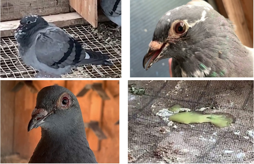
As soon as the pigeons start racing, exposure to chlamydia and other etiological agents causing ornithosis complex in the racing baskets is practically guaranteed. Clinical symptoms vary from an indistinct form, poor performance, to an acute disease which causes severe conjunctivitis, decreased appetite, respiratory tract disease, diarrhea (Figure 2), dehydration, and even death (Table 1).

Figure 2 Ornithosis complex with clinical symptoms of respiratory system and diarrhea accompanied by thin consistency of green color.
Source: Pigeonmania.28
Species |
Clinical signs |
Pigeons |
Acute Infection - anorexia, diarrhoea, conjunctivitis, rhinitis, swollen eyelids, decrease in flight performance and occasionally death (especially in young pigeons) |
Turkeys |
D serotype of the Chlamydia - Anorexia, green manure, cachexia, diarrhoea gelatinous yellow-green, low egg production, conjunctivitis, sinusitis, sneezing and mortality between 10 and 30%. |
Chickens |
Blindness, anorexia and occasionally death. |
Psittacines |
Anorexia, diarrhoea, difficulty breathing, sinusitis, conjunctivitis, yellowish droppings and, perhaps, CNS disorders. |
Ducks |
It affects mostly the young ones. Agitation, unsteady gait, conjunctivitis, serous to purulent nasal discharge and depression |
Table 1 Ornithose clinical signs in selected species of birds
Source: Modified table according to Morais et al.2
The acute form is especially widespread in young birds as well as in older individuals under stress. In some cases, conjunctivitis can lead to a secondary bacterial infection of the eye with the association of Pasteurellosis, Yersinia, Streptococci and Staphylococci which can lead to a permanently closed eyelid or even blindness.8,9 Ornithosis in pigeons can be caused only one infectious agens or can by a number of disorders which must be defined into three separate parts:
Part one is the upper respiratory such as eyes, nose, throat and sinuses. In this case, the pigeons often show only small deviations in their health status such as being overly tired after returning home, skinny dry feathers, no down feather fall, no willingness to bathe. The bath water's surface doesn't have enough white powder. In addition, there is reduced incentive for training, a blank look, blue breast muscle, feathers that stand out around the ears, rough necks, red throats, and slime in the throat. We're also referring to the "one-eye colds" phenomenon in this instance.
The second component of the middle respiratory system includes the trachea, syrinx, and bronchi. The syrinx, which creates the sound in a bird's "throat," is located about 10 inches within the body at the point where the trachea divides into the two main bronchi (Figure 3).
Part three is the deep respiratory trachea such as the lungs and air sacks. In this case, other diagnostic methods such as X-ray of the lungs and air sacs must be performed, because most problems are visible only in the upper respiratory part.3

Figure 3 Anatomical structures within an opened pigeon´s beak and throat of a bird with a respiratory infection.
Source: APC.12
Note: Bubble-like mucus is dripping from the sinus cavities through the choana and into the mouth as the front of the choana (the "slot") is swelled shut. The triangular structure in the bird's palate, which is enlarged and red, is the pharyngeal tonsil. The inflammation process has obliterated the spicules (the "fringe"), making them invisible. The bluish color of the mouth lining denotes low blood oxygen levels.
Syndrom of „one-eye colds“ in carrier pigeons
One of the most frequent causes of decline in a pigeons form and health during the racing season is the syndrome of one-eye colds. This disorder starts to appear after three or four races, especially when there is a cold and wet pre-season and postponed releases in April. Pigeons have many sinuses (useful cavities) in their heads. When the sinus lining becomes inflamed, it secretes fluid into the cavity. A network of extremely small tubes or ducts drains each sinus, but occasionally fluid accumulates more quickly than the ducts can empty. One of the major sinuses wraps around the eye and has a donut-like form. The accumulated fluid flows to the lower portion of this sinus under the effect of gravity, creating a bulge under the eye. It's interesting to note that in birds, this protrusion is only noticeable because it is made of soft tissue rather than exterior sinus bone, as it is in mammals.1
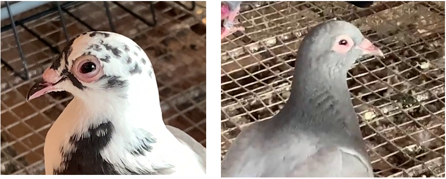
Most sinus infections are caused by either Chlamydia, Mycoplasma, or other bacteria. Chlamydia also inflames the membrane that lines the eyelids and causes conjunctivitis. The associated irritation causes excessive tearing to flow over the edge of the lid and then air dry and adhere to the feathers. The tears that are formed contain a high proportion of proteineaceous inflammatory mucus, which can cause frequent blinking movements of the eyelids. The discharge drains from the sinus through channels under the eye and deposits and stains them. It may then flow through the "slit" to the roof of the mouth or into the throat. This combination of conjunctivitis and sinusitis is what breeders call "one eye cold". Breeders often detect the first signs of infection by the presence of mucus from the oozing inflamed eyelid and bad condition of the feathers around the eye or ear.3,10 In young pigeons, symptoms are usually limited to the upper respiratory tract and the most observed symptoms are dirty nares, nasal discharge and red, watery eyes (Figure 4). However, in some pigeons, the pathogen can infect various internal organs, including the liver and spleen, as well as deeper parts of the respiratory tract, especially the air sacs. These pigeons can often be quiet, reluctant to fly, lose weight and have green sparse droppings. Pigeons with inflamed air sacs often gasp for air even during short flights around the loft, causing them to land in trees or on buildings nearby.1,11

Figure 4 Illness of the upper respiratory tract - syndrome of one-eye colds and ears without or less shiny and smooth feathers around the ears.
Source: Pigeonmania.28
In older pigeons natural immunity is higher and their reaction to this disease is different. The observed clinical symptoms are often milder than in young pigeons. Among the first symptoms of the syndrome of one-eye colds are the loss of the enthusiasm to fly, which is reflected in the deterioration of their form and poor placement in races. Pigeons that are reluctant to fly are quiet in the loft and with dry or stuck feathers (especially around the eyes and beak), they scratch their heads, wipe their eyes over the wings, sneeze, and gasp, which indicates an infection of the upper respiratory tract. Irritation of the upper respiratory tract is usually manifested by an increased frequency of sneezing more than three times in five minutes from 100 pigeons.1,3
When the beak is opened, it is possible to see inflamed tonsils, from the trachea or from the "cieft" in the middle of the oral cavity, thick white mucus can pass into the throat, which can be closed due to the swollen edges of the upper part of the oral cavity. The trachea may be red and inflamed, the beak near the opening of the nostril may be wet, the nose may be slightly coloured or there may be a slimy component on the pigeon's mucosa. The palate and the throat or muscles may be bluish in colour. Chronically infected pigeons show delayed recovery after racing and develop green droppings after stress due to liver damage. However, often the only symptoms in racing pigeons are sometimes mediocre performance and increased mortality. Pigeons with inflamed sinuses tend to cope particularly poorly on cold days with strong winds. Presumably the cold wind, irritates the already inflamed sensitive sinuses, and acts like an "ice cream cold" that makes breathing difficult.12
Diagnostic methods
It is important not to confuse symptoms with a diagnosis. Many pigeon diseases have similar symptoms. The described symptoms indicate a problem, but an accurate diagnosis can only be achieved by testing. The tests used today provide fast and accurate results that were not available to veterinarians or breeders in the past. Commonly used tests are intended for the detection of the primary agent (Chlamydia psittaci) using:
All these tests are used in veterinary practice. Less frequently used tests for detecting Chlamydia include microscopic tissue examination using special dyes. These methods focus on detecting other etiological agents of ornithosis complex by cultivating mycoplasma on special soils or by detecting the DNA mycoplasma in a sample of mucus taken from the neck using the PCR method.15,16 For bacterial diagnosis, mucus samples are inoculated on blood agar, and after cultivation, suspected colonies are re-infected on selective soils with the determination of bacterial agents using biochemical diagnostic tests17 or with matrix assisted laser desorption ionization-time of flight mass spectrometry (MALDI TOF MS).18
Treatment and eradication
Currently, treatments and strategies for reducing contamination of Chlamydial infection and other pathogens causing ornithosis complex are the best way to control the disease. Mainly, bacteria of the family Chlamydiae can survive up to 30 days in faeces, litter and cage materials or so regular cleaning of equipment and places where animals are infected is essential.19 The eradication besides the improvements to the loft with disinfection (1 to 1000 ammonium quaternary, 1 to 100 chlorophenol, 70% isopropyl alcohol and 1% Lysol)20 consist of a treatment with suitable antibiotics.1 To successfully cure pigeons, the most common antibiotics from the tetracycline series are applied, which are highly effective against Chlamydia. Doxycycline is the best because it reliably enters the cells where the bacteria are causing damage. To reduce the swelling of inflamed eyelids, terra-mycin drops are used with application twice a day for 5-7 days. However, it is very difficult to completely cure pigeons from Chlamydial infection. Even a 7-week non-stop treatment will not completely rid all pigeons of bacteria. Therefore, long-term prevention is particularly important, construction of the loft should include well-ventilated areas enabling pigeons21,22 to develop their own resistance to a relatively high degree. Another problem is that ornithosis complex is often accompanied by other diseases associated with digestive problems, or an inflamed throat or crop.3,23,24
In such a case, combined antibiotic treatment with Lincospectin for 10 days with doxycyclinone and doxycycline alone for another 4 weeks is recommended. Before starting the combined treatment, it is necessary to confirm the diagnosis by laboratory examination of samples or autopsy of dead individuals.25 One of the big problems encountered by carrier pigeon breeders is the recommended treatment period (especially for chlamydia), which should be in the range of 45-50 days, which is difficult to comply with due to the frequency of races. None of the fanciers wants to lose their pigeons unnecessarily, but they also cannot afford such a loss of time and risk of killing their competitors due to the application of long-term treatment.26 Another problem is that if breeders wanted to completely eradicate chlamydia with long-term antibiotic treatment, it is known that any immunity the pigeons have would gradually disappear. This means that if the pigeons were subsequently re-exposed to stressful situations and a new infectious dose during basketing and racing, re-infections with more severe clinical symptoms could occur.27
Whether the pigeons get problems or not after their transport and race, depends on the resistance of the pigeons concerned, the conditions of basketing and the infection pressure in the basket. It is all about balance between diesase casuing agents and the pigeon’s own resistance. It must be remembered that ornithosis is hardly ever a problem when you do not race pigeons. It will become a big problem only after the start of the racing season, which will take away the success of many breeders.
Finally, an important warning; bacteria of the family Chlamydiae can be transmitted from animals to humans. Man is mainly infected by the dispersed bacteria in the inhaled air, after faeces, urine or secretions of the respiratory system of infected animals dry. The most common manifestation is atypical pneumonia, i.e. pneumonia like cold or flu. It is therefore necessary to observe all hygienic measures when cleaning the premises of the loft and handling positive individuals.
The study was support by grant KEGA 009UVLF-4/2021: Innovation and implementation of new knowledge of scientific research and breeding practice to improve the teaching of foreign students in the subject of Animal husbandry.
Authors declare that there are no conflicts of interest.

©2023 Zigo, et al. This is an open access article distributed under the terms of the, which permits unrestricted use, distribution, and build upon your work non-commercially.