Open Access Journal of
eISSN: 2576-4578


Review Article Volume 2 Issue 5
Department of Pathology, Medical Foundation and Clinic, Nigeria
Correspondence: Wilson IB Onuigbo, Department of Pathology, Medical Foundation and Clinic, 8 Nsukka Lane, Enugu 40001, Nigeria
Received: October 30, 2018 | Published: November 16, 2018
Citation: Onuigbo WIB. Nature’s intrinsic “Erythrocyte Associated Necrosis Factor” (EANF) explains the anomalous lack of metastases in “bulky” lung cancers. Open Access J Trans Med Res. 2018;2(5):138-140. DOI: 10.15406/oajtmr.2018.02.00055
An original observation was to the effect that necrosis resulted among lung cancer cells when they were commingled with the erythrocytes in the microenvironment of the thoracic duct. Therefore, I personally named this hitherto unknown Factor as the “Erythrocyte Associated Necrosis Factor” (EANF). Can this hypothetical Factor be confirmed on the basis of its explanatory evidence? The answer was sought by considering the anomaly that “bulky” primary lung cancers scarcely metastasize. Now, it is the bulky lung cancer that develops more EANF with both the time elapsing and also with the space occupied by it. Therefore, it is because of these two productive grounds that EANF is so bounteous as to be able to face the circulating cancer cells arriving not only in the opposite lung but also in the extra thoracic sites. Accordingly, bountiful breakthrough in EANF research is called for. This will come about by
In conclusion, successful EANF research will point positively in the direction of that hoped for target therapy which could conduce to cancer cure. This particular feat will surely materialize just as when the ominous bleeding of yester years was countered successfully because of the discovery and use of the Coagulation Factor.
Keywords cancer, necrosis, natural factor, bulky cancer, replication, target therapy
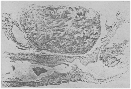
Under Professor D. F. Cappell, I went through the Scottish Pathology Residency Program in the University Department of Pathology which was housed in the Glasgow Western Infirmary. Incidentally, that Institution was acknowledged recently in the Bulletin of the History of Medicine as being second to none in Britain.1 Therein, I soon realized that the usual displaying of human parts on the Table was not sufficient for recognizing topographical relationships. Instead, I introduced the “Mono-Block Formalin-Fixation Method,” whose advantage was the precious preservation of the axial tissues from the neck to the pelvis.2 See Figure 1. Moreover, on account of serendipity, I used the Swiss-roll method to ensure that the difficult 45 cm long thoracic duct was easily prepared routinely and stained on but a single microscope slide.3 The revealed amazing panorama was strikingly that of lung cancer cells being carried more or less leisurely at the moment of death along this obviously strategic avenue. See Figure 2. In particular, I observed that, when cancer cells and red cells became commingled, necrosis occurred in this auspicious microenvironment. Accordingly, I named this extraordinary killing entity as the “Erythrocyte Associated Necrosis Factor” (EANF).4 It may be charged that it is not Necrosis per se but “coagulation.” If so, only red cells would have been seen. Moreover, if it is “inflammation,” white corpuscles alone would be involved. The thrust on EANF is on the oddity of comingling of cancer cells and red cells. Accepting this hypothesis concerning this poignant program of enduring natural events, its decoding has become necessary.

Figure 1 Specimen of mono-block formalin-fixed bodily tissues extending from above the thyroid gland to beyond the aortic bifurcation.

Figure 2 Longitudinal sections through the thoracic duct showing single cancer cells and arrowed necrotic piled up cancer cells. A metastasized lymph node is present above it.
“Bulky” lung cancers as the way forward
While preparing for the External Doctorate Degree of London University,5 I purposely consulted 67 such theses deposited throughout the University Libraries in Britain. Actually, the theses ranged from that of Aberdeen University deposited in 18926 to those of Manchester University in 1959.7,8 Incidentally, Bryson’s 1949 Cambridge Thesis9 was on the thorough analysis of 866 lung cancer necropsies. He recorded not materialized in my Glasgow series.10 See Figure 3. Yesner also experienced it.11 Accordingly, I personally named such cases as “bulky” lung cancers. Therefore, in terms of EANF, does bulkiness offer a clue? Now, it is to be expected that, with the transpiring of both time and space, EANF production in the body would increase materially. Therefore, consider this natural sequence. The bulky lung cancer is that which takes both time and space to attain its distinct dimension. On that account, the EANF produced during bulky attainment should be maximal. Since EANF is a natural element geared towards necrosis of cancer cells, it adequately assails the colonizing cells landing not only in the contralateral lung but also elsewhere in the body.
The role of the microenvironment in cancer metastasis is increasingly being recognized.12 As I see it, there is need to go even as far back as 1798, when Sir Astley Cooper13 carried out animal experiments and performed the human autopsy. In this way, he noticed in a hanged criminal that the thoracic duct can contain erythrocytes. To sum up, he surmised that this duct is very important to the “human economy.” In fact, I am persuaded that this curious conduit is naturally equipped with an almost mysterious microenvironment. Indeed, as I pointed out elsewhere,14 researches on the thoracic duct with the intravital video microscope among consenting patients15 will facilitate the retrieval of the necessary necrotic cancer cells and their commingled red cells. In all probability, the new field of translational medicine is poised to reveal the significance of EANF by retrieving the necrotic pabulum and carrying out research on it. In point of fact, the duct’s position in metastasis goes well beyond what is traditionally called “Tumor Necrosis,” seeing that this refers to the ordinary necrosis found in the primary tumor itself.16 Thus, in the words of Park’s associates,17 “Tumor necrosis was defined when there were necrotic tissues in the tumor mass at low magnification.” In contrast, the EANF is relevant because it manifests during the secondary process, i.e., “metastasis.” In other words, what goes on inside the duct itself, especially necrosis, is part and parcel of the metastasis phenomenon itself. EANF research requires the pursuit of the long known principles of science.18 In particular; the race should not be hindered by disbelief. For example, Professor Cameron’s terse truths are to be borne in mind.19 “Ideas,” he had urged, “must be sought after and cultivated.” Then, as he continued, “We must not be afraid to attempt the impossible; no device or technique that holds out the faintest hope of discovery can be put aside…” All told, I would urge, as I did recently,20 that the visionary views of the medical masters of yester years concerning the importance of tracing the very footsteps of Nature must be pursued in earnest. Thus, as Karl Pearson21 posited back in 1892, “The smallest group of facts, if properly classified, and logically dealt with, will form a stone which has its proper place in the great building of knowledge.”
In all probability, the vindication of the important role of EANF in the realm of metastases is in the horizon. In sum, when the expected breakthroughs in the field of EANF as well as target therapy become manifest, they could conduce to cancer cure just as bleeding was a killer in the old days until the Coagulation Factor was discovered.22
None.
The author declares that there is no conflict of interest.

©2018 Onuigbo. This is an open access article distributed under the terms of the, which permits unrestricted use, distribution, and build upon your work non-commercially.