Open Access Journal of
eISSN: 2576-4578


Case Report Volume 2 Issue 1
1Russian Vreden Scientific Research Institute of Traumatology and Orthopaedics of the RF Ministry of Health, Russia
2St Petersburg State University, Medical Faculty, Russia
3Pavlov First Saint Petersburg State Medical Academy, Russia
Correspondence: LN Solomin, Russian Vreden Scientific Research Institute of Traumatology and Orthopaedics of the RF Ministry of Health, Medical Faculty, St Petersburg State University
Received: November 01, 2017 | Published: February 2, 2018
Citation: Solomin LN, Korchagin KL, Tyulkin EO, et al. Ilizarov bone transport in large knee joint defect (case report). Open Access J Trans Med Res. 2018;2(1):19-23. DOI: 10.15406/oajtmr.2018.02.00029
The large knee joint defects (LKJD) are one of the indications to amputation and prosthesis. Alternative to it is reconstructive surgery based on Ilizarov method. We present the case of a female patient with LKJD 16cm and shortening of the right lower extremity 12cm, hypertrophic distraction regenerate of the right lower limb 6cm and chronic osteomyelitis of the right femur. The whole period of treatment was 67 months (5.5 years). Period of osteosynthesis (period of distraction and fixation) was 43 months (3.5 years). The complications that we faced during the treatment had no influence on good final anatomical and functional results.
Keywords: knee joint, ilizarov method, oncology, osteomyelitis, surgery
The large knee joint defects (LKJD) leads to persistent dysfunction of the lower limbs and disability.1,2 The most common reason of extended defects of the bones forming knee joint is a radical surgery for osteomyelitis, failed revision arthroplasty for oncology and Endoprosthesis replacement.3–5 In case of impossibility of an operation for the next revision endoprosthetics of knee joint, the alternative to amputation and exoprosthetics is the operation of arthrodesis of the knee joint.2,6–8 However, the presence of LKJD makes it impossible for acute shortening due to the invagination of soft tissue, vascular and neurological disorders. Therefore, for this kind of patients the reconstructive surgery based on of Ilizarov method is indicated.1,2,9
Female, 55y.o. Giant cell tumor of bone of the distal third of the right femur was diagnosed in 1985. Resection with subsequent arthroplasty was performed. In 1993 and 2002 revision arthroplasty were done. In 2003 a deep infection developed. On June 05, 2003 debridement and removal of an Endoprosthesis were performed. As a result, a large knee joint defect 30cm was formed.
In 2004 following manipulations were performed: plastic defect with vascularized fibular graft, сorticotomy of the tibia, attempt of bone defect replacement according to Ilizarov. As a result, the consolidation occurred only at the site of contact between the graft and the tibia; a hypoplastic regenerate in the middle third of the lower leg was formed. In 2006 she was admitted to the Russian Scientific Research Institute of Traumatology and Orthopedics named after R.R. Vreden with a diagnosis: 16cm of defect of the bones forming the right knee joint, 12cm of the right lower limb shortening, 6cm of hypertrophic distraction regenerate of the right tibia, chronic osteomyelitis of the right femur and lower leg, remission phase (Figure 1).
The patient refused amputation, which she has been repeatedly proposed. The first stage of treatment included the operation performed on 25 January 2006: the combined (hybrid) external fixation device (EFD) was imposed, corticotomy with osteoclasia of the middle third of the tibia, subsequent distraction to replace the defect and compression at the levels of hypertrophic regenerate and defect were performed (Figure 2).

A

B
Figure 2A & B Photos and radiographs of the patient after application of combined (hybrid) external fixation device (EFD), cortycotomy with osteoclasia of the middle third of the tibia, subsequent distraction to replace the defect and compression at the levels of the hypertrophic regenerate and the defect.
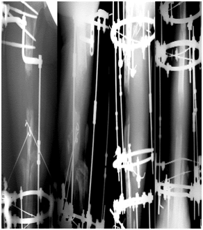
From 6 weeks beginning of treatment cortycotomy with osteoclasia of the femur and subsequent distraction to replace the defect of the bones forming the knee joint were performed. For 120 days/17 weeks of distraction received distraction regenerate at the femur with a length of 8cm, on the lower leg 13cm length (Figure 3).

A

B
Figure 3A & B Photos and radiographs of the patient after cortycotomy with osteoclasia of the femur and subsequent distraction to replace the defect of the bones forming the knee joint.
The second stage of treatment included: on 26 March 2007 [from 60 weeks beginning of treatment] an open adaptation at the junction of bone fragments of the femur and tibia was performed. For the formed equinus foot position of the right foot, Achilles tendon was lengthened, and hinges transosseous apparatus was applied (Figure 4).

A

B
Figure 4A & B Photos and radiographs of the patient after achilles tendon lengthening and application of hinge transosseous apparatus to eliminate equinoxes foot position.
The period of osteosynthesis was 30 months/132 weeks (the period of distraction of 12 months/53 weeks+fixation period of 18 months/79 weeks) 10.09.08. EFD was dismantled (Figure 5). The residual shortening of the right lower limb at that time was up to 8cm. The third stage of treatment included: On 05 October 2010 [From 248 weeks the beginning of treatment/62 months] the imposition of EFD and cortycotomy with osteoclasia of the right femur was performed.

A

B
Figure 5 Photos and radiographs of the patient in the (A) Fxation phase and (B) After dismantling of EFD.
The distraction was carried out during 110 days/16 weeks. As a result, regenerate with a length of 8 cm was formed. At the end of distraction, the correction of the mechanical axis of the lower limb with Ortho–SUV device was carried out.10 The period of osteosynthesis of this stage was 13 months/52 weeks: the distraction period was 3.5 months/14 weeks, and the fixation period was 9.5 months/38 weeks (Figure 6). Thus, the total duration of treatment was 67 months. (5.5 years). The total period of osteosynthesis (the distraction period+the fixation period) was 43 months. (3.5 years). During the treatment, due to the long period of osteosynthesis in the EFD, soft tissue inflammation repeatedly occurred in the area of the transosseous element outlet that was stopped by local application of antibiotics and bandages (category 1 complication according to Caton).11,12 The remounting of EFD was required twice due to the instability of transosseous elements (category 2 complications, according to Caton) that did not affect the treatment result.

A

B

C
Figure 6 Photos and radiographs of the patient (A) In the elimination of residual shortening and correction of the mechanical axis of the lower limb, (B) In the fixation phase, (C) After the dismantling of EFD.
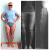
The long–term result was estimated in November 2016, 5 years after the completion of the third stage of treatment (Figure 7). The patient walks without additional means of support and uses a walking cane only for a long distance walking. The right lower limb is completely supportable, no disturbed circulations and innervations are observed. The lengths of the lower limbs are equal, the mechanical axes of the right lower limb are correct. The patient drives a car with hand control. The patient is married, brings up a child.
The frequency of infectious complications after total endoprosthetics of the knee joint, according to different authors, varies from 0.57% to 15%.9 Revision surgery, the installation of the hinge model of the Endoprosthesis – all this increases the risk of infection.13 Therefore, complications of endoprosthetics are currently the most frequent reasons for performing arthrodesis of the knee joint, when the presence of chronic osteomyelitis is a contraindication to performing revision endoprosthetics.8 The shortening of the operated limb after arthrodesis of the knee joint, after joint replacement according to different authors ranges from 1.5 to 6.4cm.8,14 In case of the large bone defect forming the knee joint after removal of cancer endoprosthesis staged reconstructive interventions are recommended.8,13,15,16 There is strong evidence that arthrodesis of the knee joint is preferable to amputation.8,15 Amputation after total endoprosthetics of the knee joint significantly reduces the quality of life, due to the impaired ability to move.17,18
However, there are few cases in the literature describing successful treatment of patients with LKJD after endoprosthetics with oncological endoprosthesis. We found only 3 publications describing the cases of substitution of extensive LKJD after a failed endoprosthesis replacement with oncological prostheses.15,16 Tokizaki cites 3 cases of treatment of patients with defects after removal of oncological prostheses with a defect value of 22–33cm, the average fixation time was 19.4 months (14.7–24.2). Kinik (2009) describes 3 cases, one of which is post–traumatic defects of 11 to 31cm, the mean fixation time was 10.1 months (7.5–15), the follow–up period was 33.6 months (25–39 months). Hatzokos describes 2 cases of substitution of extensive LKJD from which in one case the oncological prosthesis was useded after a severe fracture of the proximal tibia Schatzrer VI. The size of defects was 19 and 25cm with a fixation period of 27 and 34.7 months, respectively. Other publications18–20 describe cases of replacement of posttraumatic defects. All the authors describe complications, characteristic for external fixation – pin–tract infection11,12 and, in a number of cases, secondary foot deformity after tibia lengthening.13,15,18,19 The results of the SF–36 scale indicated that treatment significantly improved both the physical and psychological quality of life of patients.13,18
The main advantage of external fixation authors considers the simultaneous removal of limb length discrepancy and weight bearing restoration.20 The arthrodesis of the knee joint in the treatment of LKJD reduces the risk of repeated deep infection. Among the shortage of the method is an extremely long period of fixation. However, if the recommendations are observed, this does not worsen the result obtained.13 The result obtained by us on the terms of treatment, the period of fixation is not inferior to the literature data.21
The treatment of this patient with a large bone defect forming the knee joint was multi–staged, long–term and labor–intensive. However, this treatment was alternative to amputation and exoprosthetics, which the patient categorically refused. The patient is completely satisfied with the obtained results and confirms the correct choice of the therapeutic approach.
None.
The author declares no conflict of interest.

©2018 Solomin, et al. This is an open access article distributed under the terms of the, which permits unrestricted use, distribution, and build upon your work non-commercially.