Open Access Journal of
eISSN: 2576-4578


Case Report Volume 2 Issue 3
Department of Ophthalmology, University of Tunis El Manar, Tunisia
Correspondence: Daldoul Nadia, Department of Ophthalmology, University of Tunis El Manar, Habib Thameur, Tunisia
Received: December 22, 2017 | Published: May 1, 2018
Citation: Jihen B, Nadia D, Meriem O, et al. A severe case of Mediterranean spotted fever: value of ocular examination. Open Access J Trans Med Res. 2018;2(3):78-80. DOI: 10.15406/oajtmr.2018.02.00041
Mediterranean spotted fever can affect multiple organs but ocular involvement has not been frequently described. We report a case of a 42-year-old man who was hospitalized in emergency department for hemorrhagic fever. Laboratory tests showed a severe thrombocytopenia and disseminated intravascular coagulation. Few days later, he complained about bilateral blurred vision. Ocular examination found bilateral conjonctival hemorrhage, panuveitis associated with multiple retinal lesions. EN-FACE-OCT was performed. Rickettsia was confirmed by serological test. To our knowledge, this is the first description of OCT en face inflammation lesions associated with severe form of rickettsiosis.
Keywords: rickettsiosis, hemorrhagic fever, retinitis, ocular
Rickettsiosis is diseases caused by obligate intracellular bacteria. It is a zoonosis distributed throughout the world and transmitted to human by the bite of contaminated arthropods. It is endemic in the Mediterranean area and it is caused by species called Rickettsia conorii. Typical diagnosis criteria are based on the triad of high fever, general discomfort and skin rash in a patient living in or back from an endemic region for rickettsiosis. However, this triad is not always present. In fact, atypical forms can be observed with multi-organ involvement including hepatic, renal, neurological and cardiac impairment.1–4
We here in report a difficult diagnosis of rickettsiosis occuring after hemorrhagic fever. We also describe EN-FACE-OCT rickettsiosis retinal findings and emphasize the importance of ocular examination in atypical forms of rickettsiosis.
A 42-year-old patient who had not close contact with animals was hospitalized in emergency department for jaundice associated to purpura localized in the lower limbs. Physical examination revealed hepatosplenomegaly. Laboratory tests showed a severe thrombocytopenia, anemia and disseminated intravascular coagulation. Due to hemorrhagic fever etiology’s variation, absence of signs like rash, weight loss and risk factors for HIV infection, it was a challenge for doctors to make a diagnosis. The emergency physicians discussed viral etiologies such as Ebola, Hepatitis, Dengue, Yellow fever or bacterial causes especially leptospirosis. Investigations included blood culture for bacteria, Hepatitis and HIV serologies: all these latter led to negative results. The patient was treated empirically with antibiotics without any amelioration.
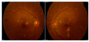
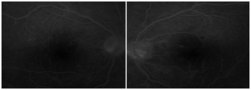
Seven days after his admission in emergency, the patient was referred to our department for blurred vision associated to ocular redness. Best corrected Snellen visual acuity was 2/10 for both eyes. A profound jaundice with conjunctival hemorrhage was found. His pupils were round and reactive, without afferent pupillary defect. The extra ocular motility was full. Also, the anterior chamber inflammation was coted 1+ and the intraocular pressure were 12mm Hg by applanation tonometry. In both eyes, there was a mild vitreous inflammation. Indirect ophthalmoscopy revealed multiple white retinal lesions in the posterior pole localized in the inner layer of retina, juxtaposed to vessels (Figure 1). These spots remained hypo or iso fluorescent in early and late frame of fluorescein angiography. We also noticed hypo fluorescent area corresponding to retinal ischemia associated to mild vascular leakage (Figure 2) & (Figure 3).


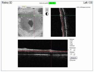
According to the inflammatory aspect of lesions, we undergo En-FACE-OCT to rule out macular edema and localize exactly retinal lesions. We found hyper reflective retinal lesions in the inner layers of the retina especially in the inner plexiform layer (Figure 4).

Leptospirosis serology, HIV, hepatitis A have returned negative. The diagnosis of rickettsial disease was confirmed by positive serology for Rickettsia conorii.
Treatment with Doxycycline 100mg twice a day was administrated for 10 days which led to systemic improvement, a visual recovery to 10/10 within two weeks and marked resolution of retinal whitening (Figure 5).
In this paper, we describe an atypical case of ocular rickettsiosis revealed by a severe hemorrhagic fever associated to thrombocytopenia and disseminated intravascular coagulation. Basically, the main etiologies of hemorrhagic fever were dominated by viral and bacterial infection, especially leptospirosis and meningococcemia. Only few cases were reported concerning association between Rickettsiosis and severe sepsis.5–9 It is due to the capacity of this small Gram negative bacterium to invade capillaries, cause endothelial injury and stimulate coagulation process which results in occlusive vasculitis. The few cases reported in literature were severe leading to fatal outcomes. The reported rate of mortality ranged between 2-3 %.10
We mention two cases: the first one about Rocky Mountain spotted fever that was associated to renal failure, coagulopathy and sepsis without history of exposure to tick bite or skin spot sign and the second one concerning a fatal spotted fever group rickettsiosis due to Rickettsia Conorii that simulated hemorrhagic viral fever.8,9 Both of these cases were diagnosed unfortunately after death according to positivity in post mortem rickettsial PCR research.8,9 For our patient, the ocular examination rectified the diagnosis. Retinitis characteristics were very suggestive of Rickettsiosis with typical lesions adjacent to retinal vessels. Moreover, other etiologies of hemorrhagic fever did not match with our ocular finding especially for leptospirosis in which membranous vitreous opacities are more common.
Ocular signs found in our patient including panuveitis associated with white retinal lesion localized in the inner layer of the retina represented a high index of suspicion for Rickettsiosis. The sequence of fluoresce in angiography showed moderate hypo or isofluorescence of the active lesion during the whole angiogram associated with vascular changes. The most common form of Rickettsiosis in the literature was represented by inner retinitis associated with moderate vitritis. This form was observed in 30% of eyes with variable topographies.10–12 The lesions varied in size, number and topography. The most frequent feature in vascular change include whether sheathing, leakage or occlusion. Basically, ocular Rickettsiosis was asymptomatic with a favorable prognosis, but atypical presentations like our case were possible.
Furthermore, we emphasize in this report the role of EN-FACE-OCT in confirming the diagnosis. EN-FACE-OCT passing through active lesions showed hyper reflectivity in the inner plexiform layer. This latter was associated to an acoustic shadowing adjacent to capillaries without subretinal exsudation. After a review of pictures of EN-FACE-OCT Atlas related to uveitis, we did not find any lesion that match with Rickettsiosis.13 As we know, the aspect of En-FACE-OCT in Rickettsiosis had never been described before. It could be considered as a reference in the characterization of this ocular disease.
Doxycycline (100 mg every 12 hours for 7 to 10 days) is the drug of choice for the treatment of rickettsial diseases. Other tetracyclines, chloramphenicol and fluoroquinolones are also effective but should be used carefully related to their potential side effects. Macrolides, including clarithromycin, azithromycin, could be used as alternative therapy in children and pregnant women.11
Clarithromycin could be an acceptable therapeutic alternative to chloramphenicol and to tetracyclines for children aged <8 years.14,15
Even though the presentation was atypical, the investigation of rickettsiosis should be kept in mind as a possible diagnosis in endemic areas. Ocular examination could have a capital contribution to diagnosis of incomplete clinical forms and help improve the prognosis. In fact, in our case, it did facilitate the case management and prescription of the appropriate treatment with systemic and ocular improvement. A careful fundus examination saves time to spend in waiting for serologic confirmation that might take 2 or 3 weeks to identify the causal agent.
Author declares no acknowledgment.
Authors declare no conflict of interest.
This article does not contain any studies with human participants or animals performed by any of the authors. Informed consent was obtained from patient included in the case.

©2018 Jihen, et al. This is an open access article distributed under the terms of the, which permits unrestricted use, distribution, and build upon your work non-commercially.