MOJ
eISSN: 2374-6920


Research Article Volume 4 Issue 3
College of Life Science, Beijing Normal University, China #both authors have equal contribution
Correspondence: Youhe Gao, Department of Biochemistry and Molecular Biology, Beijing Normal University, Gene Engineering and Biotechnology Beijing Key Laboratory, Beijing, 100875, P. R. of China, Tel +86 10 58804382
Received: October 31, 2016 | Published: November 7, 2016
Citation: Tang K, Qin W, Wang T, Gao Y. Effects of cola on the rats’ urine proteome. MOJ Proteomics Bioinform. 2016;4(3):225-227. DOI: 10.15406/mojpb.2016.04.00122
The nature of biomarker is change. Contrast to the blood which is under homeostatic controls, urine reflects changes in the body earlier and more sensitive therefore is a better biomarker source. However, the urinary proteome is affected by many factors such as diet, medication, daily activities. We studied the effect of cola intake on urinary proteome, using animal models to minimize related factors. Urine samples before and after cola treatment were collected and analyzed by SDS-PAGE and LC-MS/MS. Spectral counts was used to perform semi-quantitative analysis. In total, 371 proteins were identified with FDR<1% and at least 2 peptides. To be conservative, all of the differential proteins met the following criteria: 1); T-test values P<0.05, 2) the variation trend in all three animals was consistent, and the fold change was at least 1.5 in each rat. Twelve proteins significantly changed after cola intake, including 4 increased and 8 decreased. Six significantly changed proteins were previously annotated as urinary candidate biomarkers. Cola is a common beverage around the world; thus, this differential protein should be considered for future diseases biomarker studies.
Keywords: cola, biomarker, urinary proteome
Biomarkers are the measurable changes associated with physiological or pathophysiological process, and the most fundamental property of biomarkers is change.1 Blood is under the control of homeostasis to remain stable and balanced for protecting organs from disturbing factors, so it tends to remove changes quickly. Urine, as a filtrate of blood, without homeostatic controls, can accumulate all kinds of changes so it is a better source for biomarker discovery than blood.1,2 So far, a wide range of candidate urinary biomarkers have been reported.3
However, urinary biomarkers research can be challenging for the fact that changes in urine are very complicated. There are various elements affecting urinary proteome, diet, medication, gender, age, daily rhythms, exercise, and hormone status.4 It is difficult to determine whether potential biomarkers are truly related to diseases. We tried to use rats to study their effects one factor at a time. The results can help to annotate the potential disease biomarkers that are affected by these confounding factors.
Cola is a very common beverage in our daily life. The purpose of our experiment is to find out whether drinking cola will cause changes on urinary proteome and determine which proteins are affected.
Ethics statement and cola treatment
Four adult male Wistar rats (210-220g) were purchased from Charles River, China. All animals were kept with standard laboratory diet under controlled indoor temperature (22±1°C) and humidity (65%-70%). The experiment was approved by Institutional Animal Care Use & Welfare Committee of Institute of Basic Medical Sciences, Peking Union Medical College (Animal Welfare Assurance Number: ACUC-A02-2013-015). For these 4 rats, all drinking water was replaced with cola (commercially available sucrose-sweetened carbonated drink, Coca-Cola™, China). Before the cola treatment and at the fourth day after cola treatment, all rats were individually placed in the metabolic cages for 12hours. Then the twelve-hour urine samples were used for proteomics analysis.
Urinary proteins extraction and tryptic digestion
Urine was centrifuged at 3500g for 30min immediately after collection. Three volumes of ethanol were added after removing the pellets, and the samples were precipitated at 4°C. Then, 8M urea, 2M thiourea, 25mm dithiothreitol and 50mm Tris were used to re-dissolve the pellets. The protein concentration of each sample was measured by the Bradford protein assay. A filter-aided sample preparation method5 was used to digest the proteins in the three samples (each contained 200μg of urinary protein). Briefly, protein solution was reduced with 4.5mm DTT for 1h at 37°C, and then alkylated with 10mm IAA for 30min at room temperature in darkness. Finally, proteins were digested with trypsin (1µg/50µg protein) overnight at 37°C. The resulting peptides were desalted and then lyophilized for LC-MS/MS analysis.
LC-MS/MS analysis
The digested peptides were dissolved in 0.1% formic acid and loaded on a trap column (75µm×2cm, 3µm, C18, 100Å). The eluent was transferred to a reversed-phase analytical column (50µm×150mm, 2µm, C18, 100Å) by Thermo EASY-nLC 1200 HPLC system. Peptides were analyzed by Fusion Lumos mass spectrometer (Thermo Fisher Scientific). The Fusion Lumos was operated on data-dependent acquisition mode. Survey MS scans were acquired in the Orbitrap using a 350-1550m/z range with the resolution set to 120,000. The most intense ions per survey scan (top speed mode) were selected for collision-induced dissociation fragmentation, and the resulting fragments were analyzed in the Orbitrap. Dynamic exclusion was employed with a 30s window. Three technical replicate analyses were performed for each sample.
Protein identification and statistical analysis
The MS/MS data were processed using Mascot software (version 2.4.1, Matrix Science, London, UK) and searched against the Swiss-Prot rat database (05/03/2013; containing 9,354 sequences). Search parameters were set as follows: 10 ppm precursor mass tolerance, 0.02 Da fragment mass tolerance, two missed cleavage sites allowed in the trypsin digestion, cysteine carbamidomethylation as fixed modification, oxidation (M) as variable modifications. The Mascot results were filtered and validated by Scaffold (version 4.0.1, Proteome Software Inc., Portland, OR, USA). Peptide identifications were accepted if they were detected with 90.0% probability and an FDR less than 0.1% by the Scaffold local FDR algorithm. Protein identifications were accepted if they were detected with FDR less than 1%and contained at least 2 identified peptides. Spectral counting was used to compare protein abundance between different experimental groups. To be conservative, all of the differential proteins met the following criteria: 1); T-test values P<0.05, 2) the variation trend in all three animals was consistent, and the fold change was at least 1.5 in each rat.
SDS-PAGE analysis of urinary proteins before and after cola treatment
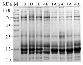
25μg urinary proteins were separated using 12% SDS-PAGE gel. There were consistent changes before and after cola treatment. As is shown in Figure 1, during 55-100kDa and 130-180kDa, the bands are lighter after drinking cola, while about 15kDa, the bands are darker, indicating that drinking cola will have an obvious impact on the rat's urinary proteome (Figure 1).

Urine proteome changes identified by LC-MS/MS
To investigate the effects of cola intake on the urinary proteome, the urinary proteomes from three rats before and after cola intake were analyzed by LC-MS/MS. Six samples three rats (three samples from cola treatment before, and three after treatment) were analyzed by UPLC coupled with Fusion Lumos. In total, 371 proteins were identified with FDR<1% and at least 2 peptides (Supporting Information Tables S1). For the total difference in spectra after the cola treatment, spectral counting was used to perform semi-quantitative analysis. The details of criteria are described in Materials and methods.
Twelve proteins were significantly altered after cola intake (Table 1). Four proteins increased and 8 decreased, 9 proteins have human orthologs. All significant changed proteins were searched against the Urinary Protein Biomarker Database.3 Six significantly changed proteins were previously annotated as urinary candidate biomarkers (Table 1). Cola is a common beverage around the world; thus, these protein changes should be considered when researching diseases biomarkers.
ID |
Protein Name |
MW |
PSM |
PSM |
Fold Change |
Change Tendency |
Human Ortholog |
Diseases |
|
||||||||
1 |
2 |
3 |
1 |
2 |
3 |
Rat1 |
Rat2 |
Rat3 |
|
|
|
||||||
P20759 |
Ig gamma-1 chain C region |
36 |
0 |
2 |
3 |
5 |
6 |
7 |
∞ |
3 |
2.3 |
UP |
NO |
None |
|
||
P02625 |
Parvalbumin alpha |
12 |
0 |
1 |
0 |
2 |
2 |
3 |
∞ |
2 |
∞ |
UP |
P20472 |
Compound-induced skeletal muscle toxicity6 |
|
||
P42854 |
Regenerating islet-derived protein 3-gamma |
19 |
13 |
15 |
16 |
49 |
87 |
53 |
3.8 |
5.8 |
3.3 |
UP |
NO |
Membranous nephropathy7 |
|
||
P27590 |
Uromodulin |
71 |
77 |
74 |
63 |
149 |
140 |
164 |
1.9 |
1.9 |
2.6 |
UP |
P07911 |
Sepsis-induced acute renal failure8 |
|
||
P07897 |
Aggrecan core protein |
221 |
3 |
6 |
4 |
2 |
1 |
1 |
0.7 |
0.2 |
0.3 |
DOWN |
NO |
None |
|
||
O35112 |
CD166 antigen |
65 |
3 |
4 |
3 |
2 |
1 |
2 |
0.7 |
0.3 |
0.7 |
DOWN |
Q13740 |
Type 1 diabetes11 |
|
||
P42123 |
L-lactate dehydrogenase B chain |
37 |
5 |
4 |
6 |
3 |
1 |
2 |
0.6 |
0.3 |
0.3 |
DOWN |
P07195 |
None |
|
||
P01026 |
Complement C3 |
186 |
14 |
10 |
17 |
7 |
3 |
8 |
0.5 |
0.3 |
0.5 |
DOWN |
P01024 |
None |
|
||
P07150 |
Annexin A1 |
39 |
4 |
7 |
5 |
2 |
1 |
1 |
0.5 |
0.1 |
0.2 |
DOWN |
P04083 |
Glomerular injury12 |
|
||
P50399 |
Rab GDP dissociation inhibitor beta |
51 |
4 |
4 |
3 |
2 |
2 |
2 |
0.5 |
0.5 |
0.7 |
DOWN |
P50395 |
None |
|
||
B5DFC9 |
Nidogen-2 |
153 |
3 |
4 |
4 |
1 |
1 |
2 |
0.3 |
0.3 |
0.5 |
DOWN |
Q14112 |
None |
|
||
Q9JJ40 |
Na(+)/H(+) exchange regulatory cofactor NHE-RF3 |
57 |
13 |
12 |
15 |
4 |
7 |
8 |
0.3 |
0.6 |
0.7 |
DOWN |
Q5T2W1 |
|
|||
Table 1 Details of changed urinary proteins before and after drinking cola
Thanks to the Australian Proteome Analysis Facility (APAF) for funding this research.
The author declares no conflict of interest.

©2016 Tang, et al. This is an open access article distributed under the terms of the, which permits unrestricted use, distribution, and build upon your work non-commercially.