MOJ
eISSN: 2381-179X


Case Report Volume 8 Issue 4
Oral and Maxillofacial Department, King Fahd General Hospital, Saudi Arabia
Correspondence: Norah Alqahtani, Resident of Oral and Maxillofacial, King Fahd General Hospital, KSA, Saudi Arabia,
Received: June 21, 2018 | Published: July 5, 2018
Citation: Moussa K, Alqahtani N, Othaman AA, et al. Large maxillary adenomatoid odontogenic tumor: case report. MOJ Clin Med Case Rep. 2018;8(4):145-147. DOI: 10.15406/mojcr.2018.08.00261
Background: The Adenomatoid Odontogenic Tumor is a cystic hamartoma arising from odontogenic epithelium. The clinical course of the lesion is slow and remains clinically unnoticeable for a long time. It is primarily found in young female patients, located more frequently in the maxilla in most cases associated with an unerupted permanent tooth. Treatment is surgical excision, and the prognosis is excellent. Recurrence is rare. The aim of the case is to highlight the scarcity of Adenomatoid Odontogenic Tumor and to consider it among the differential diagnosis of any Maxillofacial cystic lesion.
Case presentation: A 13- year-old Saudi young man, with no past medical history, was admitted for Maxillofacial mass. The clinical manifestation as well as the imaging findings were toward Adenomatoid Odontogenic Tumor. Our patient underwent surgical excision of mass. The patient had no recurrence at one years of follow-up.
Conclusion: The unique radiological manifestations of the lesion helped in the diagnosis, but accurate histological diagnosis is mandatory to avoid unnecessary mutilating surgery. It was managed conservatively with no evidence of recurrence. However, a close follow-up is mandatory.
Keywords:adenomatoid odontogenic tumor, adenomatoid odontogenic cyst, AOT, dentigerous cyst, odontogenic myxoma.
The adenomatoid odontogenic tumour is a cystic hamartoma arising from odontogenic epithelium. It will characteristically have a lumen lined by epithelium from which proliferations fill much and sometimes all the lumen space, then mimicking a solid tumour.1 (AOT) was first elucidated by Driebaldt in 1907 as “pseudo-adenoameloblastoma” and later as “adenomatoid odontogenic tumour.”2 The World Health Organization (WHO) in 2005 defined AOT as a tumour composed of odontogenic epithelium, presenting a variety of histo-architectural patterns, embedded in mature connective tissue stroma, and characterized by slow and progressive growth. 3 This lesion is known to be allied with unerupted canines and lateral incisors. The clinical course of the lesion is slow and remains clinically unnoticeable for a long time. The deformity produced by this lesion manifests as displacement of adjoining teeth and an obvious expansion of the surrounding bone.4,5 Sometimes, it may be also refined as “two-thirds tumour” because:
Two-third occurrence in maxilla
Two-third female preponderance
Two-third association with unerupted tooth
A13-Year-old male patient reported with the chief complaint of the painless swelling in the maxillary anterior region in the last 5 Months. The swelling was not associated with any history of trauma, pain or discharge.
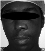
On extra oral examination revealed facial asymmetry with obliteration of naso-labial fold. On inspection, a diffuse swelling was seen on the left middle third of the face measuring approximately 3x3cm2 in size, extending anteroposteriorly from the ala of the nose to 2 cm in front of the preauricular region and superioinferiorly from the infraorbital rim up to 0.5 cm above the corner of the mouth (Figure 1). On palpation, the swelling was firm-to-hard in consistency and not tender. Intraorally, an oval-shaped swelling was seen on the buccal aspect extending anteroposteriorly from the mesial aspect of upper left lateral incisor to the distal aspect of left upper 1st molar, measuring approximately 3x2cm2 in size, superioinferiorly from the marginal gingival to upper buccal vestibule. Obliteration of buccal vestibule was present, missing upper left canine was also noted.
Panoramic Radiographic was obtained shows a large unilocular radiolucency on the left side of the maxilla, extending from tooth #22 to #26. Unerupted tooth #23 was present inside the lesion. Computed Tomography (Figure 2) (Figure 3) of facial bone was obtained shows well circumscribed unilocular radiolucency and radiolucency involves the crown of unerupted tooth #23. Based on the history and clinical features and radiographic evaluation dentigerous cyst and adenomatoid odontogenic tumour was made as provisional diagnosis. Other strictly radiolucent lesions worthy of consideration are keratocystic odontogenic tumor, ameloblastic fibroma, odontogenic myxoma, or central giant cell tumour as well as unicystic ameloblastoma which is common in this age.
The surgical excision of the cyst was done under general anaesthesia along with the removal of the unerupted canine and sent for the histopathology lab which confirm the diagnosis of (AOT) (Figure 4-6). The patient has been under follow-up for one year and was rehabilitated with fixed prosthesis.

Figure 1 Volume increase observed in the left genian region. Lesion was approximately 4 mm in diameter as well as indurated and asymptomatic.
The adenomatoid odontogenic tumor is a cystic hamartoma arising from odontogenic epithelium. Various synonyms have been used for this tumour. These are adenoameloblastoma, ameloblastic adenomatoid tumour, adamantinoma, epithelioma adamantinum or teratomatous odontoma.8 They mostly occur in young people, peak incidence is in the second decade of life, uncommon in patients above 30 years of age. The incidence of female to male ratio is 1.8:1. It is predominant in the anterior region of the jaws. They are frequently encountered in the maxilla than in the mandible, with a ratio of 2:1 and are most commonly associated with impacted teeth. Canines are the most common tooth associated with AOT.9 AOT is divided into two variants: central and peripheral. Further, the central variant is divided into two subtypes: follicular and extra-follicular. The central variant occurs at rate of 97.2%, out of which, 73.0% are follicular. The follicular variant is three times more common than the extra-follicular type. The follicular variant is diagnosed earlier in life (mean age 17 years) than the extra-follicular type (mean age 24 years); 53.1% of all variants occur within the teen years (13-19 years). A study showed that follicular AOT was associated with one embedded tooth in 93.2% of the cases. Maxillary permanent canines account for 41.7% and all four canines for 60.1% of AOT-associated embedded teeth.5
The follicular variant is associated with unerupted tooth surrounding the crowns and is attached to the necks of the tooth.10 The extra-follicular variant is not associated with an impacted tooth, superimposed over the roots of the teeth. A peripheral variant occurs over the gingiva and looks as a gingival growth.10,11
Panoramic Radiograph does give a comprehensive view of the lesion, but a multi-slice CT scan provides the extension of the lesion in all directions besides showing dental involvement, as in this case it revealed intracapsular presence of canine tooth which couldn't hence be saved. It also showed involvement of the maxillary sinus and aided in treatment planning. Quality and quantity of the scattered radiopaque foci can also be well appreciated on the CT scan. CT scan provides proper extensions of the AOT, reveals proximity to the vital structures which aid in the preoperative evaluation, provisional diagnosis, planning and execution of the surgical procedure.
On Histologic and immune-histochemical evidences are present for the follicular variant showing that they originate from the reduced enamel epithelium of the dental follicle.12 Origin of the extra-follicular variant is less clear. Philipsen et al.,13 suggest that all the AOT variants show identical histology, have a common origin and implicate the dental lamina or its remnants. Macroscopically, AOT is surrounded by a connective tissue capsule. When dissected, it may appear as a solid mass, as seen in our case14 (Figure 5). The AOT has a distinctive histopathologic appearance, i.e. spindle-shaped epithelial cells are arranged in the form of sheets, strands or whorled masses surrounded by connective tissue stroma. These epithelial cells may form rosette-like structures with a central space, which may be empty or contain an eosinophilic amyloid- like material. The characteristic feature of AOT is the presence of duct-like structures that are surrounded by columnar cells with nuclei oriented away from the lumen. These structures are not true ducts, and no glandular elements are present in the tumour. Small foci of calci cations are scattered throughout the tumour. It may also present in the lumen of the structures.13–16
In the treatment of AOT, most authors now believe that complete enucleation with long-term observation is the preferable treatment. Since all variants of AOT show identical, benign biological behavior and since most of the cases are well encapsulated, conservative surgical enucleation has proved to be the treatment of choice with rare recurrence.15,17,18 Though AOT is not known to recur, but Toida et al. reported two cases, with recurrences, and one of them had intracranial extension.19 Philipsen & Reichart15 reported recurrence in only three cases out of 750 studied.15 Long-term follow-up is thus necessary to observe the complete filling of the residual bony defect, fate of the involved tooth/teeth and recurrences if any. Guided tissue regeneration in conjunction with bone grafting can be used as an aid for rapid filling of large defects surrounding teeth created by odontogenic tumors.
In our case, the lesion being larger and follow-up being longer, we could observe the corrected deformity and the bony cavity had healed optimally in the absence of any adjunctive therapy. The patient was referred for prosthodontist for dental rehabilitation and subsequent implant after patient exceed 18 years.
We thank Dr. Nadia Enani Consultant Maxillofacial Histopathologist Department, King Fahad General Hospital, KSA.
Author declares that there is no conflict of interests.

©2018 Moussa, et al. This is an open access article distributed under the terms of the, which permits unrestricted use, distribution, and build upon your work non-commercially.