MOJ
eISSN: 2381-179X


Case Report Volume 8 Issue 2
1College of Medicine,University of Illinois at Chicago, USA
2Department of Pathology, University of Illinois at Chicago, USA
3Department of Dermatology, Cook County Health and Hospitals System, USA
Correspondence: Kayla St Claire, University of Illinois at Chicago, College of Medicine, 1740 Taylor Street, Room 3132W, Chicago IL 60612, USA
Received: March 14, 2018 | Published: March 27, 2018
Citation: Claire KS, Braniecki M, Sachdev D,et al. Primary varicella presenting with coexistent features of PLEVA. MOJ Clin Med Case Rep. 2018;8(2):67–68 . DOI: 10.15406/mojcr.2018.08.00242
We present a case of varicella-associated PLEVA in a 27-year-old man to raise awareness of this rare cutaneous inflammatory disorder. Classic clinical presentation, histology, etiology, and management will be discussed.
Keywords: pityriasis lichenoides et varioliformis acuta (PLEVA), mucha-habermann disease, varicella-zoster virus (VZV)
Pityriasis lichenoides et varioliformis acuta (PLEVA), otherwise known as Mucha-Habermann disease, is a rare cutaneous inflammatory disorder that presents as an acute eruption of multiple hemorrhagic papules with eventual necrosis, ulceration, and crusting. PLEVA most commonly occurs on the trunk, extremities, and flexural areas and typically affects children and young adults. Although the pathogenesis is unknown, literature postulates that PLEVA represents a hypersensitivity reaction to infectious agents given its association with recent or concurrent infections. We herein report a case of varicella-associated PLEVA in a 27-year-old man.
A 27-year-old male was admitted for fever and rash that started 2 days prior to admission. The rash began on his face and subsequently had spread to the rest of his body including skin folds, oral mucosa, and the genital region. He also had suffered flu-like symptoms including sore throat, generalized weakness, and myalgias, which started 1 week prior to the rash. The patient had no significant past medical history. He is a Mexican immigrant with an unclear history of prior chickenpox exposure and vaccinations.
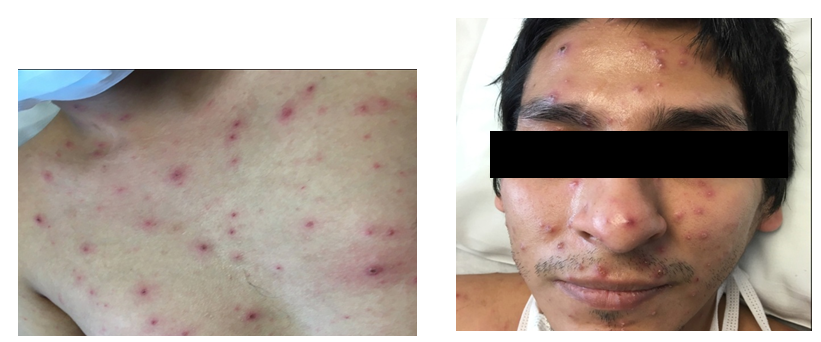
Skin examination revealed diffuse hemorrhagic vesiculopustules in different stages of healing involving the trunk, extremities, and face (Figure 1A)(Figure 1B). A skin punch biopsy from the right leg showed features of PLEVA with a lymphocytic infiltrate along the dermoepidermal junction, RBC extravasation, and epidermal necrotic keratinocytes. There was marked subepidermal edema with inflammatory cells and interface dermatitis, which extended down the hair follicle with intraepithelial apoptotic cells, but distinct herpetic viral cytopathy was not appreciated (Figure 2)(Figure 3). However, varicella-zoster virus (VZV) was detected by PCR from this skin site. The patient had elevated IgM titer to varicella, but no detected IgG antibodies to VZV. The patient was then diagnosed with primary varicella (chicken pox) and discharged on Valacyclovir 1gm TID for 7 days. At follow-up visit one-week later lesions were resolving.

Pityriasis lichenoides et varioliformis acuta (PLEVA), otherwise known as Mucha-Habermann disease, is a clinical subtype of pityriasis lichenoides (PL). PLEVA is characterized by erythematous macules that quickly evolve into papules. These papules often have a central punctum that become vesiculo-pustular. They can undergo hemorrhagic necrosis and become ulcerated with overlying red-brown crusts.1 These lesions usually heal in weeks to months, however, hyper/hypopigmented scars may remain.2 PLEVA typically presents on the trunk, extremities, and flexural areas.3 Associated symptoms may include burning, pruritus, and mild constitutional symptoms. PLEVA most often occurs in childhood with a slight male predominance.
The diagnosis of PLEVA is established clinically and by histopathology. The classic histopathologic findings of PLEVA include: confluent parakeratosis, basal layer vacuolization, exocytosis of lymphoid cells, and a perivascular lymphoid infiltrate in the dermis.4 Immunohistochemistry can also aid in the diagnosis of PLEVA and help to exclude closely resembling alternative diagnoses. For instance, since the inflammatory infiltrate in PLEVA is primarily composed of CD8+T cells, CD30 stains are usually negative unlike lymphomatoid papulosis.5 Multiple inflammatory and infectious disorders share clinical features with PLEVA. Common differential diagnoses include lymphomatoid papulosis, arthropod bite reactions, secondary syphilis, erythema multiforme, viral exanthems, vasculitis, and drug eruptions.4
The etiology of PLEVA is unknown, although it has been suggested that foreign antigens such as infectious agents are the pathogenic trigger.6 In 2003, Boralevi et al suggested a potential role of VZV in the pathogenesis of PLEVA. He demonstrated that 8 out of 13 patients with histologically confirmed PLEVA had also revealed positive VZV PCR in skin samples.6 Other infectious agents that have been implicated in PLEVA include cytomegalovirus (CMV), group A Streptococcus, Epstein-Barr virus, and HIV. Under this same theory with extrinsic antigens being the trigger, case reports have mentioned the occurrence of PLEVA following drugs (estrogen-progesterone and chemotherapeutics) and vaccines (measles, mumps, rubella (MMR) and influenza).1,7 Another hypothesis describes PLEVA as a T cell lymphoproliferative disorder due to identification of T cell clones (associated with Hodgkin’s lymphoma and lymphomatoid papulosis).1
Clinical management of PLEVA is difficult due to uncertain etiology; however, the disease is benign and tends to be self-limited. When PLEVA does persist, first line therapies include UVB phototherapy and systemic antibiotics, with tetracyclines being used in adults and erythromycin for children.1 Second-line therapies include methotrexate for refractory disease and systemic corticosteroids for severe cases.1
We share this case to raise awareness of VZV and its coexistence of PLEVA, which can lead to a better understanding in the pathogenesis of PLEVA.
None.
The author declares there is no conflict of interest.

©2018 Claire, et al. This is an open access article distributed under the terms of the, which permits unrestricted use, distribution, and build upon your work non-commercially.