MOJ
eISSN: 2471-139X


Calabash chalk (CC) is consumed mostly by pregnant women in the tropics to reduce salivation and by some other persons for pleasure. The aim of this study was to examine the effects of CC on the cerebellum of gestating wistar rats. Thirty (30) female and ten (10) male rats weighing 150-230g were used for this study. Following vaginal smear, they were mated at proestrous with the males. The day after mating was designated as day 0 of gestation. The pregnant rats were randomly divided into five groups (Control, All trimester, 1st trimester, 2nd trimester and 3rd trimester) of six each and were given 0.5ml aqueous solution of CC containing 40mg/ml for differentdays during their gestation period except the control group which were given 0.5ml of distilled water. The all trimester group were given the aqueous solution throughout their gestation period, 1st trimester 1-6days, 2nd trimester 7-12days and 3rd trimester 13-18days. On gestational day 20, the rats in all the groups were sacrificed, their cerebellum excised and fixed in 10% formol calcium and were routinely processed for histological analysis. There were no significant differences (P<0.05) that existed in the body weight between control and experimental groups. Histological analysis of the maternal cerebellum using Haematoxylin and eosin, Cresyl Fast Violet and Feulgen staining techniques showed changes in the cytoarchitecture of the cerebellar layers and hyperplasia of the granule cells in the experimental groups.
CC causes histomorphological and cellular density change in the cerebellum of gestating wistar rats which can lead to cerebellar dysfunction such as inability to maintain equilibrium or clumsy movement.
Keywords: calabash chalk, cerebellum, haematoxylin and eosin, cresyl fast violet and feulgen
CC, calabash chalk; ALL TRIM, all trimester; 1ST TRIM, first trimester; 2ND TRIM, second trimester; 3RD TRIM, third trimester; Al2Si2O5OH4,10 aluminum silicate hydroxide; CFV, cresyl fast violet
The practice of eating soil, clay, or chalk, a form of pica called Geophagy1occurs deliberately or accidental.2 It is seen in most parts of the world3 but more extensive in the tropics.4 Reports show that it occurs with animals, as well as humans, in both sexes, and in all races;5 in UK it is associated with immigrants from South Asia6‒8 and West African.9,10
Geophagy, although often seen in rural or preindustrial settings among children and pregnant women11 is not limited to any particular age or time.12 Clay consumption has been reported to be associated with pregnancy, and some women eat clay to eliminate nausea.13 Report says that this process may also result in the absorption of dangerous toxins and eggs of parasites that may have been passed in animal faeces.14 Anaemia, which is a deficiency of red blood cell and haemoglobin (oxygen-carrying pigment in the blood), as well as Ascaris lumbricoides infection, are some of the other factors associated with geophagy.14
CC (calabash clay, Calabar stones) is also called poto, la craie or Argile by the Francophones; Nzu by the Igbos, and Ndom by the Efiks/Ibibios of Nigeria. It is also referred to as Mabele by the Lingala of Congo. Although commercially available in blocks/pellets, it is also sold in powdered form.15 CC is made up of aluminum silicate hydroxide, which is a well-known member of the kaolin clay group, with the formula: Al2Si2O5OH4.10 Other substances which could be poisonous to the body have also been reported to be present in clay.10 These include metals, metalloids, and persistent organic pollutants.10 The metals include iron, aluminium, potassium, titanium, barium, chromium, zinc, manganese, nickel, rubidium, copper, and tin with the metalloids being lead the mean concentration of approximately 40mg/kg and arsenic.10,15‒17
Various organic pollutants have been reported to be present in clay/CC. Such organic pollutants include alpha lindane, endrin, endosulphan II, and P, PI-dichloro diphenyl dichloroethane (DDD).10 The European Union recommends that the highest concentration of lead permitted in specific foods should not exceed 1mg/kg (Commission Regulation, 2001).18 However, lead levels in CC have been reported in the range of 10-50mg/kg.10
Due to the high consumption of calabash chalk by pregnant and breastfeeding women as remedy for morning sickness, this study investigated the possible effects of calabash chalk on the cerebellum of gestating wistar rats.
Thirty (30) female wistar rats and ten (10) male rats weighing 150-220g and 190-230g respectively were used for the experiment. Blocks of salted calabash chalk were grounded into powder using a manually operated grinder. A 40g sample of the powder was dissolved in 1000ml of distilled water.
The solution was left for 24hours and was filtered. The filtrate was stored in a plastic container and kept in the refrigerator. Vaginal smear test was carried out prior to mating; this was done to know the phase of estrous cycle of female rats before introducing the male rats.19 The following day was taken as day zero (0) of pregnancy.19
The pregnant rats were randomly divided into five groups of six each and were given 0.5ml aqueous solution of CC containing 40mg/ml for different days during their gestation period except the control group which were given 0.5ml of distilled water. The all trimester group were given the aqueous solution throughout their gestation period, 1st trimester day 1-6, 2nd trimester day 7-12 and 3rd trimester day 13-18. On gestational day 20, the rats in all the groups were weighed and sacrificed by cervical dislocation; the maternal brains were then excised and weighed. Tissues for histological studies were fixed in 10% formol calcium and processed using Hematoxylin and Eosin, Cresyl fast violet (CFV) for demonstration of Nissl substance and Feulgen stain for demonstration of DNA.
Data obtained were analyzed using ANOVA t-test (LSD) and the data presented as Mean ± standard error of mean (SEM), with confidence interval at 95%.
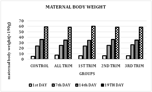
During gestation there were no significant difference (P<0.05) that existed in the maternal body weight, maternal cerebellar weight between the control and the experimental groups. However, there were weekly body weight gains among all the groups as the experimental groups were exposed to the same 0.5ml aqueous solution of CC.
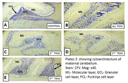
Histological observation using CFV and Feulgen stain showed destruction of normal cytoarchitecture of the maternal cerebelli and hyperplasia of the granule cells as well as decreased staining intensity for Nissl cells and the nuclei, compared with animals in the control groups (Figure 1).
Histological observations
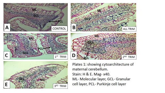
In Group A, the molecular layer was distinct and the granular layer is densely packed with small dendritic cells. The cells that form the Purkinje layer are visible. The demarcation between the molecular and granular layer can be seen. The molecular and granular layer of Groups B, C and D are less stained, the cells that form the Purkinje layer are not visible. In Group E, the demarcation between the molecular and granular layer can be seen showing the cerebellar layers (Figures 2-4).

Figure 1 Bar chart showing maternal body weight of wistar rats during gestation.
ALL TRIM, all trimester; 1ST TRIM, first trimester; 2ND TRIM, second trimester; 3RD TRIM, trimester There was no significant difference between the control and the treated groups (P<0.05).

Figure 2 Showing cytoarchitecture of maternal cerebellum. Stain: H& E. Mag-×40.
ML, molecular layer; GCL, granular cell layer; PCL, purkinje cell layer

Figure 4 Showing cytoarchitecture of maternal cerebellum. Stain, CFV. Mag-×40.
ML, molecular layer; GCL, granular cell layer; PCL, purkinje cell layer
The granule cells in Group A are deeply stained. Group B shows deeply stained and large granular cells. Group C shows less stained and destructive cells. Group D and E shows deeply stained and fewer cells when compared with control (Figure 5).
Group A: showed distinct cerebellar layers. The molecular layer appeared more distinct with a visible higher distribution of cells; the granular layer becomes denser while the purkinje layers are also visible. The demarcation between the molecular and granular layer are visible. The Purkinje layers are not visible in the treated groups. There was apparent distortion in the regular arrangement of the treated group compared to control. The granular layers are not visible in group C (Figure 6).
Group A showed deeply stained granular cells. Group C, D and E showed hyperplastic granular cells while Group B showed vacuolation of the neurons (Figure 7).
Different adverse reports have been ascribed to calabash chalk’s consumption. Higher cellular population, hypertrophied pyramidal cells, and vacuolation in the cerebral cortex,11 reduction in erythrocyte indices,15 risk to the mental development of the developing unborn babies and breast feeding infants,16 liver damage, gastrointestinal alterations,13,20 and demineralization of the femur bone.21
During gestation there were no significant differences (P<0.05) that existed in body weight between control and the experimented groups. However, there were weekly body weight gains among all the groups as the experimented groups were exposed to the same 0.5ml aqueous solution of calabash chalk. This result indicates that the rats may have had the same baseline age, growth rate and pregnancy rate. Thus, the chalk may not have had effect on the body weight of the gestating rat. This indicates that administration of calabash chalk may not always affect the body weight of the maternal. This agrees with the work of11 which showed no differences in body weight gain by the dams between the control and the treated groups following an exposure to calabash chalk.
The histological analysis of the maternal cerebellum using haematoxylin and eosin, cresyl fast violet and feulgen staining techniques showed distortion of the external germinal layer (EGL), vacuolation of the Purkinje cells, changes in the cytoarchitecture of the cerebellar layers and hyperplasia of the granule cells in the treated groups of the maternal. These changes will lead to cerebellar dysfunction. This correlates with the work of11 which showed section of the cerebral cortex with a higher cellular population, hypertrophied pyramidal cells, and vacuolation in the treatment groups which indicated that calabash chalk may have anxiolytic effect especially at high dose in the light and dark field but not in the open field and can stimulate cellular changes in the maternal cerebral cortex. Ali and colleagues deduced that high consumption of yoyo cleanser bitters for long period of time will have a significant effect on the histology of the cerebellum of wistar rats which leads to hypertrophied granular layer of the cerebellum with a corresponding increase in granular cells.22 Lorton and Anderson in their study on quantitative histological examination and Golgi analysis of the cerebellum, revealed a number of alterations in the lead treated rats.23 Lead exposure resulted in a significant decrease in the molecular layer width (72%). The granule cell density was depressed in the lead exposed rats, despite the observation that the granule cell layer width did not differ significantly.23 This agrees with the findings of our study since calash chalk contains above the recommended doses/levels of the aforementioned element/compounds required for human consumption.
The components of the calabash chalk such as lead, arsenic, aluminum, and kaolin are known to cross the blood-brain barrier and to cause different effects in different parts of the brain. Neurons are not known to proliferate when traumatized; therefore the consumption of 0.5ml aqueous solution of calabash chalk containing 40mg/ml during gestation should be discourage because it causes changes in the maternal cerebellar layers which could lead to cerebellar dysfunctions such as inability to maintain equilibrium or clumsy movement.
None.
Author declares that there is no conflict of interest.

© . This is an open access article distributed under the terms of the, which permits unrestricted use, distribution, and build upon your work non-commercially.