MOJ
eISSN: 2471-139X


Case Report Volume 3 Issue 4
1Department of Anatomy, Federal University of Pernambuco, Brazil
2Catholic University of Pernambuco, Anatomy Laboratory, Brazil
3Department of Prosthetics and Oral Facial Surgery, Federal University of Pernambuco, Brazil
Correspondence: Carla Cabral dos Santos Accioly Lins, Federal University of Pernambuco, Centre for Biological Sciences, Department of Anatomy, Av Prof Moraes Rego s/n, 50730-000 Recife, Brazil, Tel +55 81 21268555, Fax +55 81 2126 8554
Received: February 20, 2017 | Published: April 6, 2017
Citation: Accioly Lins CCS, Corrêa DIM, Lima R, et al. Anatomical study and variation of the anterior belly of digastric muscle: case report. MOJ Anat Physiol. 2017;3(4):118–120. DOI: 10.15406/mojap.2017.03.00099
The digastric muscle is one of the supra hyoid muscles that that assist in the chewing movements, usually consisting of two bellies: one anterior and other posterior, united by an intermediate tendon. This study aimed to report the presence of a previous accessory belly in digastric. The dissection was performed on a human body, adult, male, fixed and preserved in 10% formaldehyde solution, which belongs to the Anatomy Department of the equity of Federal University of Pernambuco, Recife, Brazil. During dissection of the neck in the submental region found a unilateral variation of the left digastric. The accessory belly was inserted into the digastric fossa of the contra lateral jaw, and It was thought attached to previous bellies and later by an intermediate tendon attached to the lower horn of the hyoid bone, and the length 33mm and width of 6mm.Thus, we emphasize the importance of knowledge of the diversity of morphological arrangements as a way to differentiate multiple variations, thus facilitating diagnostic and surgical procedures in the anterior neck.
Keywords: anatomic variation, digastric muscle, dissection
The digastric muscle development takes place in the pharyngeal arches at the beginning of the fourth week of intrauterine period, when neural crest cells migrate to the future of the head and neck area.1‒3 It consists of two bellies, anterior and posterior, which has different embryological origin.4,5 The anterior belly develops from the first pharyngeal archor mandibular arch, and is innervated by mylohyoid nerve, a trigeminal branch; and the posterior belly that develops the second pharyngeal archor hyoid arch, which is innervated by the seventh cranial nerve, also known as facial nerve.5‒7
The anterior belly goes through posterolateral inferiorly intermediate tendon to the digastric fossa on the inner face of the jaw (insert), and the posterior belly passes anteroinferiorly on the medial side of the mastoid incisure of the temporal bone (source) to the intermediate tendon, i.e, both linked by a single tendon attached to a lower horn of the hyoid bone.3,5,8,9
The digastric muscle is one of the suprahyoid muscles that assist in the chewing movements, but the main muscles of mastication are: masseter, temporal and medial and lateral pterygoid.7 Anterior bellies, right and left, of the digastric muscles divide the region between the hyoid bone and mandible in two triangles: the submental and submandibular. The submental triangle is a unique area, which demarcates the lower limit, the base, the body of the hyoid bone and laterally the anterior bellies of digastric muscles. The submandibular triangle is a paired area between the lower border of the mandible and the anterior and posterior bellies of digastrics.9‒11 Knowing that this region has a number of important anatomical structures, and a good knowledge of anatomy helps surgical procedures involving the region, we aimed to describe this case report of morphological variation of the anterior belly of the digastric.
During dissection routine in anatomy laboratories of the Federal University of Pernambuco, Recife, Brazil, in a human body, adult male, fixed and preserved in 10% formaldehyde solution, an anatomical variation of the digastric muscle was observed. Initially, it was taken the skin, adipose tissue, muscle sheath and the platysma muscle was folded out. In submental region found a left unilateral variation of the digastric. This piece was photographed using a Sony Cyber-shot DSC-W30 camera, and the bellies were marked and measured using a caliper PA, with an accuracy of 0.05mm.
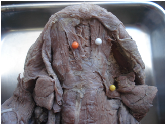
In this study, the anterior belly of the digastric muscle has an accessory bell that arises from the contralateral digastric fossa of the mandible, which it is attached to the anterior e posterior bellies by an intermediate tendon fixed to the lower horn of the hyoid bone (Figure 1). It has a length of 33mm and width 6mm, and other surrounding structures were normal in all respects, on both sides (Figure 2).

Figure 1 Region suprahyoid identifying the digastric muscle.
![]() Anterior Belly Accessory;
Anterior Belly Accessory;
![]() Anterior belly;
Anterior belly;
![]() Posterior belly.
Posterior belly.
The variations of the anterior belly of the digastric can be discovered during routine dissection, imaging techniques and or surgical procedures. Some cases were identified and reported from the past centuries to the present day.9,12 The literature describes a frequency of 0.2%, which is extremely rare13 however, some researchers have found described this variation in more than 50% of the cases,9,14 most often unilaterally, but may also appear bilaterally.2,11,14 It is believed that the diversity of morphological arrangements for digastric muscle, whether resulting from the union of two different muscles embryological which are connected by an intermediate tendon forming the anterior and posterior bellies.8
Several authors have reported the anatomical variations of the digastric, describing them according to location, trajectory, innervation and composition.3 In a study by Liquidate et al.,7 the authors described four cases of anterior belly variations. They observed that in both cases the accessory belly was located on the right side one was inserted in to mylohyoidraphe, and the other had path similar to the one shown in this study. The other two cases the variation was bilateral with presence of two wombs accessories, in which both originated medially to the digastric muscle digastric fossa in the jaw, but one was inserted in the contralateral intermediate tendon and the other in mylohyoidraphe.
Çelik et al.15 reported a variation of the anterior belly of the left digastric, which featured four different ipsilateral inserts and four branches of the mylohyoid nerve. These muscle bundles were attached to the intermediate tendon and continued with the posterior belly. As well, Aktekin et al.16 described a rare event bilateral and symmetrical variation of the anterior belly of the digastric muscle in the form of cruz. Others authors report the presence of a previous bilateral accessory belly originating in the intermediate tendon and inserted in mylohyoidraphe.3
Besides the antinodes of the number of variations described in most of the articles, there are other variations of the digastric muscle also cited in the literature, such as rare cases in which the anterior belly of the muscle is innervated not only by the trigeminal nerve, as normally, but also by a branch of the facial nerve.17,18
Unilateral anatomical variations may be more relevant when considering the clinical situation, since in some cases they may be responsible for the asymmetry in the front of the neck, or even during the movement of the floor of the mouth or temporomandibular joint.2 These types of asymmetry can lead to functional wear, and can be confused with lymph nodes, benign or malignant cervical masses in clinical tests, imaging (MRI, CT, ultrasound) and surgical procedures.7,19‒22
This study aimed to emphasize the need for a better understanding of normal anatomy in order to differentiate the multiple anatomical variations of the anterior belly of the digastric muscle, thus facilitating diagnostic and surgical procedures on the anterior area of the neck.
None.
Author declares that there is no conflict of interest.

©2017 Accioly, et al. This is an open access article distributed under the terms of the, which permits unrestricted use, distribution, and build upon your work non-commercially.