MOJ
eISSN: 2471-139X


Research Article Volume 3 Issue 3
1Department of Morphology and Postgraduate Applied Health Science Program, Federal University of Sergipe (UFS), Brazil
2Dentistry student, Federal University of Sergipe (UFS), Brazil
3Medical Student, University Center of Volta Redonda (UNIFOA), Brazil
4Titular Professor, Medical School, Tiradentes University (UNIT), Brazil
Correspondence: Jose Aderval Aragao, Associate Professor of the Federal University of Sergipe and Titular Professor of the Medical School of the Tiradentes University, Rua Aloisio Campos 500, Bairro Atalaia, Aracaju, Sergipe, Brazil, CEP 49035020
Received: November 01, 2016 | Published: March 17, 2017
Citation: Aragão JA, Silveira MPM, Cisneiros de Oliveira LC, et al. Determination of sexual dimorphism from the area of the triangle. MOJ Anat Physiol. 2017;3(3):79–82. DOI: 10.15406/mojap.2017.03.00091
A triangular area can be formed from lines connecting the infraorbital foramina (IOFs) and the nasion to each other. Measurements on this triangle may help to determine sexual dimorphism in human skulls. The aim of this study was to determine sexual dimorphism in Brazilian skulls from measurements of the area of the triangle formed between lines joining the IOFs to the nasion. 242 human skulls belonging to the Study and Research Center for Anatomy and Forensic Anthropology of Tiradentes University. It was observed that out of the 242 skulls examined, 148 were male and 94 were female, with ages ranging from 18 to 91years (mean: 57years). In the male skulls, the distance between the IOFs ranged from 53 to 58.8mm (mean: 56mm). In the female skulls, it ranged from 50.7 to 56.0mm (mean: 53.7mm). In the male skulls, the mean distance from the right IOF (RIOF) to the nasion ranged from 44.1 to 49.0mm (mean: 46.2mm) and from the left IOF (LIOF) to the nasion, 44 to 48mm (mean: 46.0mm). In the female skulls, the distance from the RIOF to the nasion ranged from 42 to 47mm (mean: 44.7mm) and from the LIOF to the nasion, 41.8 to 46.1mm (mean: 44mm). The area of the triangle formed by the lines connecting the IOFs to the nasion in the male skulls ranged from 944.6 to 1105.3mm² (mean: 1023.9mm²). In the female skulls, it ranged from 853.8 to 1025.3mm² (mean: 946.5mm²). A sexual dimorphism occurred in all measures and area that formed the triangle between the infraorbital foramen and the nasion that were larger in males.
Keywords: anthropometry, anatomy, sexual dimorphism, infraorbital foramen, nasion
N, nasion; RIOF, right infraorbital foramen; LIOF, left infraorbital foramen; DRIOF, distance from the right infraorbital foramen to the nasion; DLIOFN, distance from the left infraorbital foramen to the nasion; DIOF, distance between the infraorbital foramina; PIOFN, perimeter formed by lines between the infraorbital foramina and the nasion; ATIOFN, area of the triangle formed by lines between the infraorbital foramina and the nasion
The human skull is considered to be the second most useful indicator for determining an individual’s sex, after the pelvis.1 It is also responsible for furnishing important data for identifying the sex of living individuals and even cadavers.2,3 For forensic experts to perform these tasks, precise methods and techniques are required.4,5
Male skulls are generally formed by coarser or rougher structures because of the presence of stronger muscle insertions. Moreover, they are also larger than female skulls.4,5
Two methods are used in the process of determining individuals’ sex from parts of the craniofacial skeleton: the qualitative or morphological method and the quantitative or metric method. Most studies have used the morphological method, in which characteristics such as the frontal sinuses, teeth, glabellae, bone thickness, eyebrow ridge thickness and chin shape have been studied.5 On the other hand, the quantitative method makes use of measurements between pre-established points on the skull. This method, which is considered to possibly be more efficient, has contributed greatly towards determining individuals’ sex.2,6
Most authors in the literature on skull measurements from quantitative or metric variables have used samples from other countries (i.e. not from Brazil).6‒8 This places limitations on the applicability of these studies to the Brazilian population, given that without knowledge of the parameters for measurements on Brazilian skulls, researchers are obliged to use international tables. This could lead to uncertainty regarding the results.2
Therefore, the present study had the objective of determining sexual dimorphism in Brazilian skulls from measurement of the area of the triangle formed by lines between the infraorbital foramina (IOFs) and the nasion.
In this study, 242 dry human skulls were used: 148 male skulls and 94 female skulls, ranging in age from 18 to 91years. The skulls had been obtained in accordance with law no. 8501 of 1992, which deals with use of unclaimed cadavers for use in studies and research. These skulls belong to the Study and Research Center for Anatomy and Forensic Anthropology (CEPAAF) of Tiradentes University.
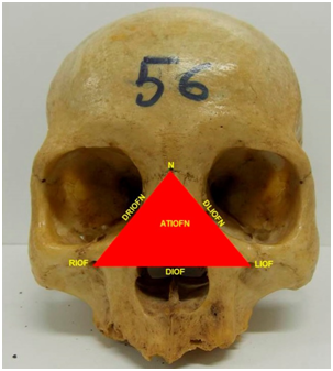
This was a morphometric study in which the following were measured: distance of the right infraorbital foramen to the nasion (DRIOFN), distance of the left infraorbital foramen to the nasion (DLIOFN), distance between the infraorbital foramina and perimeter and area of the triangle formed bylines between the infraorbital foramina and the nasion (Figure 1). The linear measurements were made with the aid of digital calipers with accuracy to the nearest 0.01mm.

Figure 1 Triangle formed by the infraorbital foramina and the nasion.
N: Nasion; RIOF: Right Infraorbital Foramen; LIOF: Left Infraorbital Foramen; DRIOFN: Distance From the Right Infraorbital Foramen to the Nasion; DLIOFN: Distance From the Left Infraorbital Foramen to the Nasion; DIOF: Distance Between the Infraorbital Foramina; ATIOFN: Area of the Triangle Formed by Lines Between the Infraorbital Foramina and the Nasion
The data were analyzed both descriptively and analytically. The normality of distribution of the numerical variables was assessed by means of the Shapiro-Wilk test. Variables that met the assumption of normal distribution were presented as the mean (M) and 95% confidence interval. Spearman’s linear correlation test was applied and the values obtained were categorized as follows: 0 to 0.39 as weak correlation; 0.40 to 0.69 as moderate correlation; and 0.70 to 1.00 as strong correlation. To compare morphometric values according to sex, the Mann-Whitney test was applied. To evaluate multivariate differences we apply MANOVA and Lambda Wilk’s Test. The statistical significance level was stipulated as 5% (p≤0.05). For all the analyses, the Statistical Package for the Social Sciences (SPSS 15.0) software was used.
In the sample of 148 male skulls and 94 female skulls, there were no abnormalities with regard to age. The median age of the male skulls was 57years (41.75-70) and the median of the female skulls was 56.50years (41.50-70). There was no statistically significant difference in age between the sexes of the skulls (p=0.887).
In relation to sex, there were statistically significant differences in the measurements of DIOF, DRIOFN, DLIOFN and the perimeter and area of the triangle delimited by the infraorbital foramina and the nasion (Table 1) and a multivariate difference in measurements.
Variables |
Male M (IC95%) |
Female M (IC95%) |
p |
DIOF |
56.0 (53.0-58.8) |
53.7 (50.7-56.0) |
< 0.001* |
DRIOFN |
46.2 (44.1-49.0) |
44.7 (42.0-47.0) |
< 0.001* |
DLIOFN |
46.0 (44.0-48.0) |
44.0 (41.8-46.1) |
< 0.001* |
PIOFN |
147.7 (142.5-154.0) |
142.5 (135.0-147.6) |
< 0.001* |
ATIOFN |
1023.9 (944.6-1105.3) |
946.5 (853.8-1025.3) |
< 0.001* |
Table 1 Morphometric variables in relation to sex.
DIOF: Distance Between the Infraorbital Foramina; DRIOFN: Distance from the Right Infraorbital Foramen to the Nasion; DLIOFN: Distance from the Left Infraorbital Foramen to the Nasion; PIOFN: Perimeter formed by Lines between the Infraorbital Foramina and the Nasion; ATIOFN: Area of the Triangle Formed by Lines Between the Infraorbital Foramina and the Nasion Mann-Whitney test; *Significance (p ≤ 0.05).MANOVA: Lambda Wilk’s=0.82, F_94,148=10.12, p<0,001.
The results from the comparisons of measurements between the sexes showed that all of them were greater in the male skulls. It could be seen from Spearman’s linear correlation that all the correlations between age and all the measurements of distance, perimeter and area were weak or insignificant (Table 2).
DIOF |
DRIOFN |
DLIOFN |
PIOFN |
ATIOFN |
|
Age |
0.049 |
-0.002 |
-0.013 |
0.011 |
0.009 |
Rho (P-Value) |
-0.447 |
-0.978 |
-0.836 |
-0.868 |
-0.892 |
Table 2 Correlation analysis between age and all the measurements of distance, perimeter and area
DIOF: Distance between the Infraorbital Foramina; DRIOFN: Distance from the Right Infraorbital Foramen to the Nasion; DLIOFN: Distance from the Left Infraorbital Foramen to the Nasion; PIOFN: Perimeter Formed by Lines Between the Infraorbital Foramina and the Nasion; ATIOFN: Area of the Triangle Formed by Lines Between the Infraorbital Foramina and the Nasion
Determination of sex is considered to be one of the main pillars of human identification protocols. This can be achieved through analysis of cranial measurements and visual evaluation of the skull, among other means.1,4,5,7,9‒14 Because craniofacial structures are seldom destroyed, they have been widely used by several authors for determining sex.1‒6,9,10,15,16 The quantitative method was used in the present study to determine sex. Some authors have considered that this method is more reliable when pre-established points are used and have accepted that qualitative method are subjective and unsafe.2‒6,11 Almeida Junior et al.17 using the logistic regression technique, for this type of study, obtained a score of 71.8%.
We did not find any use of the triangular area formed between the infraorbital foramina and the nasion for determining sexual dimorphism, in the literature. Almeida Junior et al.5 conducted a similar study, but they used the distance from the infraorbital foramina to the prostion as craniometric reference points. These authors found that the mean distance between the infraorbital foramina was 60.94mm for male skulls and 58.25mm for female skulls. Regarding the distance from the infraorbital foramina to the prostion, the mean was 31.30mm for male skulls and 29.47mm for female skulls. Regarding the area formed by lines between the infraorbital foramina and the prostion, the mean was 956.68mm² for male skulls and 860, 46mm² for female skulls. In the present study, the mean DIOF was 56.0mm for the male skulls and 53.7mm for the female skulls. Regarding the area of the triangle, the mean obtained was 1023.9mm² for the male skulls and 946.5mm² for the female skulls. For all the variables studied, the means were greater in the male skulls.
Badam et al.9 and Patil & Mody3 analyzed linear skull measurements obtained via lateral cephalometry. In both studies, the nasion was used as a reference point in order to establish and compare results between the sexes. Like in the present study, the values obtained were greater in the male skulls. In our study, comparisons of the DIOF, DRIOFN, DLIOFN and the perimeter and area of the triangle formed by lines between the infraorbital foramina and the nasion, in relation to the sexes, all showed statistically significant differences, such that all of the measurements were greater in the male skulls. This may be explicable in terms of the anatomical differences between male and female skulls.
Studies involving visual analyses on skulls have indicated differences between the sexes. For example, prominences, bone crests and other anatomical abnormalities are more perceptible in male skulls than in female skulls.2,4,5,9,16‒19 However, according to Badam et al.,9 Patil & Mody,3 Shearer et al.13 and Spradley & Jantz,1 metric analysis makes it possible to obtain results that are more objective, especially for researchers with little experience of determining individuals’ sex. Moreover, this facilitates the statistical analysis on the sample and comparison with other studies.
Many studies conducted around the world have used quantitative variables in their analyses.7,10,12,13,16‒18,20‒23 In these analyses, the researchers only used international data because they did not have any measurements on Brazilian skulls. According to Almeida Junior et al.,4 Almeida Junior et al.,5 Bigoni et al.,17 Francesquini Junior et al.,2 Franklin et al.,18 Gapert et al.,7 Kimmerle et al.11 and Kruger et al.,12 this may compromise the analysis because of the possibility that a variety of factors such as climate, local geography, diet, socioeconomic conditions and quality of life might interfere with defining an individual’s sex. In this regard, Almeida Junior et al.4 accepted that difficulties in the process of determining sex might arise through variations in measurements and morphology found in different populations. However, authors such as Ferreira et al.,24 Almeida Junior et al.25 and Lima et al.26 believe in the possibility of using their findings, in Brazilian skulls, in services of Forensic Dentistry and in the practical field of Forensic Anthropology.
The area of the triangle formed by lines between the infraorbital foramina and the nasion, and also all the other variables, were greater in the male skulls, with a statistically significant difference. This means that anatomical characteristics are of fundamental importance in distinguishing between the sexes, given that factors such as climate, local geography, diet, socioeconomic condition and quality of life may interfere in defining an individual’s sex.
Author declares that there is no conflict of interest.

©2017 Aragão, et al. This is an open access article distributed under the terms of the, which permits unrestricted use, distribution, and build upon your work non-commercially.