MOJ
eISSN: 2471-139X


Mini Review Volume 3 Issue 2
Department of Cell Biology, Universidad Nacional Autonoma de México (UNAM), Mexico
Correspondence: Luis F Jimenez-Garcia, Laboratorio de Nanobiologia Celular Departmento de Biologia Celular, Universidad Nacional Autonoma de Mexico (UNAM), Circuito Exterior, C.U., 04510, Cd. Mx, Mexico, Tel (55) 56 22 49 88, Fax (55) 56 22 48 28
Received: October 14, 2016 | Published: February 15, 2017
Citation: Jiménez-García LF, Lara-Martínez R, Garza-Melchor RE, et al. The nucleolus of Giardia lamblia. MOJ Anat Physiol. 2017;3(2):41–43. DOI: 10.15406/mojap.2017.03.00083
The nucleolus is the major site of ribosome biogenesis in eukaryotes. Until recently, G. Lamblia was considered the only eukaryote lacking nucleoli. Recently, light and electron microscopy cytochemical techniques have been used to demonstrate the presence of nucleoli in the interphase nucleus of G. Lamblia. Here we review the work made during the last few years on the nucleolus of G. Lamblia in interphase and cell division. This mini review indicates that nucleoli are present in G. Lamblia interphase cell nuclei. Also, the persistence of nucleoli during cell division was documented more recently. Therefore, microscopical anatomy of G. Lamblia should include the presence of authentic morphological nucleoli.
Keywords: G. Lamblia, G. Duodenalis, nucleolar, golgi apparatus, endoplasmic reticulum, hybridization, nuclei, mitosomes, flagella, ribonucleoprotein
AgNOR, silver staining for nucleolar organizer region; NOR, nucleolar organizer region; Rrna, ribosomal rna; TEM, transmission electron microscopy
G.Lamblia, also known as G. Intestinalis or G. DSuodenalis, is a unicellular parasite infecting human large intestine and causing severe diarrhea.1‒6 According to B. Ford, it was first observed by Antony van Leeuwenhoeck back in 1681,7 as very small organisms, with a large body and a flattish belly with little paws and stir movements. G. Lamblia was later described taxonomically.8
G.Lamblia also belongs to early divergent lineages of eukaryotes as revealed by protein or nucleic acids sequence analysis,#ref99‒11 although reduced processes also may have occurred.12 Studies on the cell biology of G. Lamblia have described several organelles as nuclei, mitosomes, flagella, several elements of cytoskeleton, but definite proof for the presence of Golgi apparatus or endoplasmic reticulum have not been documented.1‒6 For a long time G. Lamblia was known as the only eukaryote lacking nucleoli. Since the detailed analysis searching for nucleoli failed to show the presence of nucleoli in Giardia even by using immunocytochemistry and in situ hybridization combined with the use of confocal microscopy,13 it was asked whether classical cytochemical staining as silver staining for nucleolar organizer would produce better contrast than fluorescent techniques.
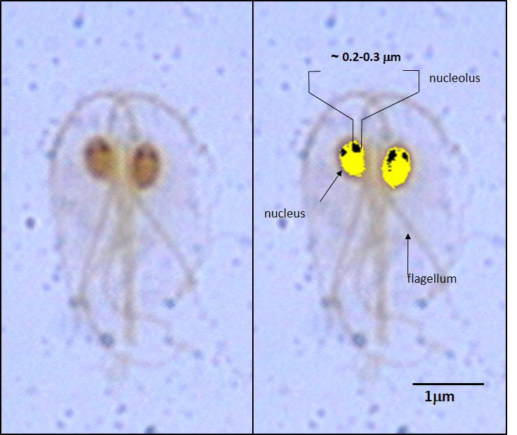
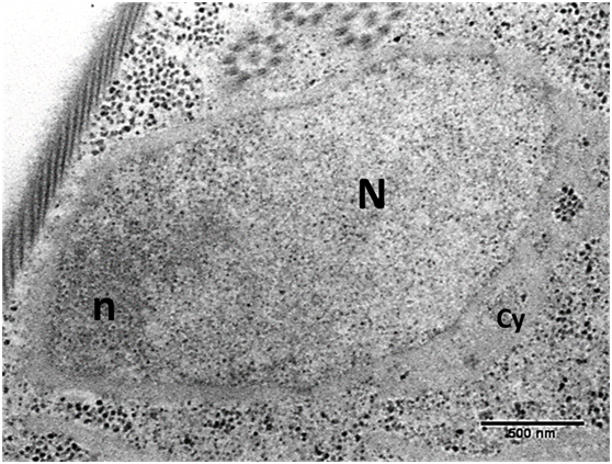
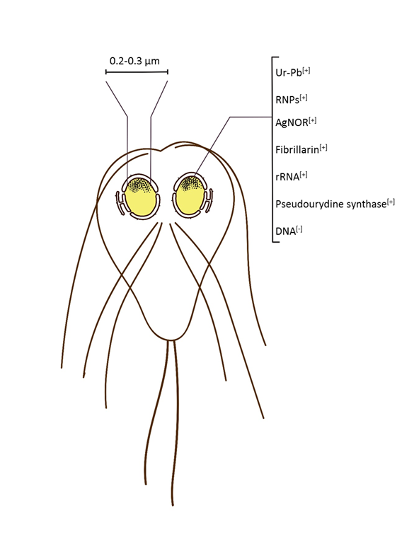
The first evidence for the presence of nucleoli in G. Duodenalis, G. Intestinalis or G. Lamblia was obtained after using silver staining for nucleolar organizer regions (AgNOR) for light microscopy. Nucleolar material was observed as small fiber-granular areas of impregnation around 0.3µm large (Figure 1).14 Then, transmission electron microscopy (TEM) confirmed this result. In fact, standard technique for TEM produced results indicating an intranuclear electron dense material at the periphery of nuclei (Figure 2), that in addition was positive to silver staining for NOR, for RNA using the EDTA method for RNA, silver staining for NOR at the EM level followed by immunoelectron microscopy for fibrillarin and high resolution in situ hybridization for rRNA. Analysis of expression of transfected rRNA-pseudouridine synthase (CBF5) and confocal microscopy confirmed these results. Later on, additional light and electron microscopy techniques also revealed the presence of nucleoli (Figure 3).15 Microscopical anatomy of G. Lamblia therefore, includes the presence of nucleoli.4‒6,16

Figure 1

Figure 2 Transmission electron microscopy of G. lamblia nucleolus. Nucleolus (n) is present at the periphery within the cell nucleus (N).
Cy: Cytoplasm; n: Nucleolus; N: Cell Nucleus

Figure 3 Drawing of G. lamblia showing the nucleolus in both nuclei of the cell summarizing the different nucleolar markers tested. Ur-Pb [+], positive in samples contrasted with uranyl acetate for standard transmission electron microscopy; RNPs [+], positive for the EDTA regressive staining for ribonucleoproteins; AgNOR [+], positive for silver staining for nucleolar organizer region; Fibrillarin [+], positive for immunoelectron microscopy for nucleolar protein fibrillarin; rRNA [+], positive for high resolution in situ hybridization for rRNA; pseudourydine synthase [+], transfection for expression of this protein; DNA [-], negative for immunoelectron microscopy for DNA.
More recently, it was reported further confirmation of the presence of nucleoli in G. Lamblia in interphase and also the persistence of this organelle through cell division,17 therefore indicating that the process of nucleolar biogenesis does not include the formation of the so called pre nucleolar bodies, as in other eukaryotes.18
G. Lamblia displays the smallest nucleoli so far described in nature. It is about 0.2-0.3µm large, which is also about the resolving power for standard light microscopy. The reason of why it was not observed before, may have to do with its size and the use of fluorescent probes that, even they are so specific, however they do not display the highest contrast. On the other hand, silver staining for nucleolar organizer offers the highest contrast when analyzing by bright field light microscopy. The first observations suggested that a fine study had to be made with TEM. The best fixation techniques and further cytochemical, immunocyto chemical and in situ hybridization techniques at the ultrastructure level confirmed that nucleolar material was present in G. Lamblia. A fibrous and granular component characterizes an intranuclear region. Further analysis with transfection of the gene for the rRNA-pseudourydine synthase or fibrillarin also confirmed the presence of a nucleolar domain within the nuclei of G. Lamblia.14
The results mentioned for the presence of this nuclear organelle in such early divergent eukaryote organism,14 suggest that nucleoli may be considered a sine qua non structure for eukaryotes, further suggesting that the origin of nucleoli predates the origin of nuclei. In addition, the persistence of nucleolar material during cell division,17 may also indicate that nucleoli are active through cell division, an aspect that have to be explored. In fact, the role of active nucleolus or nucleolar material during cell division may confer some advantages in such environments as stomach and intestine. Recent results also have been observed in other unicellular parasites as Trypanosoma cruzi.19
Nucleoli are present in both interphase nuclei of the unicellular parasite G. Lamblia. We found in current literature that G. Lamblia nucleolus was about 0.2µm in size, is present as peripheral fibro-granular ribonucleoprotein material, is positive to several nucleolar markers and it is negative for DNA presence. Morphological and cytochemical evidence indicate that nucleoli are persistent during the cell cycle.
Supported by CONACyT (180835), PAPIIT-DGAPA-UNAM (IN217917). María Teresa Jiménez made the drawing of Figure 3.
Author declares that there is no conflict of interest.

©2017 Jiménez-García, et al. This is an open access article distributed under the terms of the, which permits unrestricted use, distribution, and build upon your work non-commercially.