MOJ
eISSN: 2471-139X


Research Article Volume 2 Issue 3
Department of Anatomy, Yenepoya Medical College, Yenepoya Universiy, India
Correspondence: Venkatesh Kamath, Department of Anatomy, Yenepoya Medical College, Yenepoya Universiy, Deralakatte, Mangalore, Karnataka, India, Tel +919164190758
Received: February 16, 2016 | Published: April 23, 2016
Citation: Kamath V, Asif M, Bhat S, et al. A prototype of digitalized anatomy museum with a scientific design. MOJ Anat Physiol. 2016;2(3):69–75. DOI: 10.15406/mojap.2016.02.00046
Anatomy museums display the intricacy of human anatomy to medical students and public. This study aims to create a prototype of a digitalized twenty first century museum. The prototype was conceived after a study on sixteen renowned anatomy museums in India. The museums have evolved over the centuries from an institute housing a collection of models, to one with formalin preserved specimens and plastinates. The modern anatomy museums are digitalized and use audiovisual aids and electronic screens to express anatomy. The advances in science must be utilized in medical education. The future generations live in world that is completely digitalized. Hence, modern innovations such as computer, internet, Wi-Fi, electronic screens, projectors, 3-D viewing capabilities, 3-D holograms and anatomical mannequins, must be used to teach anatomy in anatomy museums. It is also essential to incorporate museum visit, as a part of regular curriculum, in schools and in undergraduate medical education. An anatomy museum is not meant only for display of specimens. It has to be designed, to function as an institute where active learning can take place, thus including the museum in the mainstream of medical education. The future of anatomy will certainly be more digitalized. However, the traditional techniques of model making, dissection and plastination will always have a special place in all the anatomy museums for time immemorial.
Keywords: anatomy museums, digitalization, formalin preservation, medical education, models, plastinates
A museum may be defined as an institute that displays a collection of artifacts of scientific or artistic significance to a specific group of visitors.1 Anatomy museums play a dominant role in educating the community regarding the intricacy and structural organization of the human body.2 The concept of anatomy museum was first conceived by the Edinburg Surgeons during 1700-1763 A.D. A cabinet containing anatomical pictures, books and few specimens was first prepared and this was called the “cabinet of curiosities”.3 Early museums were institutes housing a simple collection of models of anatomical specimens. The models were made of diverse materials like wood, clay, wax and ivory.4 The discovery of formalin in 1859 by Alexander Mikhailovich Butlerov and its subsequent isolation in 1868 by August Wilhelm Von Hoffmann was a great milestone in anatomical history, as it completely changed the manner in which anatomy would be preserved for future generations.5 Since 1868, the anatomy museums have evolved significantly with the invention of novel museums techniques. Plastination was invented in 1978 at the University of Heidelberg by Gunther von Hagens.6 There is a significant transition in the nature of collections exhibited in Anatomy museums. The museums have metamorphosed substantially, from a collection of models and art to express anatomy in eighteenth century, to the use of formalin preserved specimens in nineteenth century, to plastinates in late twentieth century.7 The La Specola Collection,8 the Hunterian museum,9 Museum of the Royal College of Surgeons of Edinburg,10 Anatomy Museum of Queen’s University of Belfast11 and the Pedro Ara Anatomy Museum12 are some renowned international anatomy museums. The modern museums are digitalized and use audio-visual aids to display anatomy. This study is an original research on Indian museums, which aims to collect information on all aspects of museum. This information has been used to design a prototype of a digitalized anatomy museum, which would benefit both the medical students and the public. The authors believe that an anatomy museum is not meant only for display of specimens. It has to be designed, to function as an institute where active learning can take place, thus including the museum in the mainstream of medical education.
Sixteen Indian Anatomy museums were chosen from sixteen reputed medical colleges in India. Permission was obtained from the concerned authorities in all medical colleges for conducting the study. All aspects of the museum were studied. This included specimen preparation and preservation techniques, mounting, labeling, jars, display, novel inventions like plastination, museum maintenance, staff, types of lightings and racks, passage, interior and digitalization. The manner in which information was stored in the computers and made accessible to students was observed. In this article, the authors would like to restrict the discussion to designing a prototype of a digitalized museum.
In this article only specific observations are discussed, with an objective of creating a sophisticated museum that can educate both medical students and public.
Skilled dissection
It is fine dissection, performed with skilled hands under expert guidance, which creates an impact on the museum visitors. The clarity with which the anatomical intricacy is preserved during dissection determines the specimen quality. It was observed; that the best among the museums visited had technicians, specially trained in the art of dissection, preservation and display. They were trained under the guidance of eminent anatomists of the University to create models, mummies, corrosion casts and palatinates that unveiled the splendor of human anatomy. A finely dissected specimen of large intestine with the arterial circle of Drummond is shown in Figure 1.
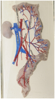
Figure 1 Skilled dissection is the foremost requisite for a good museum. The brances of inferior mesentric artery and the arterial circle of Drummond are finely dissected by the anatomists.
Courtesy, Kasturba Medical College Anatomy Museum, Manipal
Specimen preparation
It is essential to select a fresh specimen for dissection. Following appropriate ethical clearance, a fresh autopsy specimen or a well embalmed specimen must be procured. The edges must be cut sharply with fine instruments. The size of the specimen to be displayed and the manner in which it is to be removed from the body must be planned. The method of fixation is also important and it is essential to follow steps described by standard literatures. A well prepared specimen showing the brain and spinal cord of a 4month old fetus is shown in Figure 2. The figure clearly depicts the fact that planning and visualization play a significant role in specimen preparation. Bones can be prepared either by putrefaction method or by autoclaving.
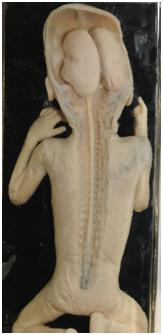
Figure 2 Planning and visualization play a significant role in specimen preparation, as observed in this specimen of brain and spinal cord of a 4 month old fetus.
Courtesy, Kasturba Medical College Anatomy Museum, Manipal
Specimen preservation
It is essential to preserve the specimens, in a preservative that maintains the color of the specimen, with minimal fading over the years. It was observed that the museums with good display use Pulvertaft-Kaiserling’s technique for preservation. The technique uses three solutions, first for fixation, second for color restoration and a third for color maintenance. Pulvertaft in 1936 discovered that addition of sodium hydrogen sulphite prevented fading of specimen.13 Perspex jars are preferred for preservation, as they are clear, light weight, tough, durable, easy to cut and have ideal optical properties.
Mounting of specimen and labeling
A lot of emphasis is laid to mounting a specimen and labeling. The technique of mounting can be very creative and certainly creates an impact on the visitors. A well mounted neuroanatomy specimen of basal ganglia is shown in Figure 3.
Structuring the museum
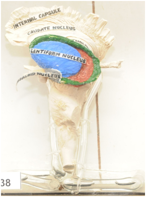
Figure 3 Innovative mounting enhances the display. A well mounted specimen of basal ganglia.
Courtesy, Kasturba Medical College Anatomy Museum, Manipal
A prototype of a digitalized anatomy museum designed by the authors is shown in Figure 3. The first section on the left hand of the entrance displays portraits of eminent anatomists and their contributions. The other sections mentioned below, must be constructed in an order, as depicted in the prototype. Each section must be provided with audio-visual aids, a computer and a few study tables and chairs for students to contemplate and study the section. The last section on the right hand side of the entrance will be for collecting feedback and for providing museum related literature to the visitors. The concept here is to incorporate the advances in science, computer, internet and audio-visual aids in each and every section of anatomy museum. Trained guides can be used to explain the videos to the visitors. It is also essential to keep different visiting hours for public, the school children and the medical students.
Anatomists’ contributions: Portraits of eminent anatomists can be displayed in this section. The contributions of anatomists can be explained using audio-visual aids. The evolution of dissection techniques over the centuries can be described using artistic sketches and videos.
Evolution of species: Anatomical knowledge is always incomplete without understanding evolution of species. The theories describing evolution can be displayed using charts and translite boards. Similarly, overhead projectors can be used to provide a brief session on evolution.
Comparative anatomy: Human anatomy can be compared with anatomy of other species. Both the gross and osteology parts of different species can be compared. Example: A stomach, a liver or a heart of different species can be compared. Comparative osteology can be displayed as shown in Figure 4.
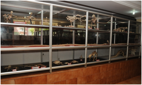
Figure 4 A Comparative osteology section enables comparison of osteology of different species.
Courtesy, Kasturba Medical College Anatomy Museum, Manipal
Developmental anatomy: Development of organs and organ systems can be described using embryology models. Diverse materials like plaster of Paris, wood, rubber, clay, acrylic and glass can be used to prepare the models. Specimens of human fetus at different stages of gestation can be displayed. A specimen of a three month old fetus in an amniotic sac is shown in Figure 5. Fetal skeleton can be stained with alizarin staining. Developmental anomalies can be displayed as shown in Figure 6. Audio-visual aids such as electronic screens and projectors can be used to describe the development of fetus. Special videos must be designed for public, school children and medical students.
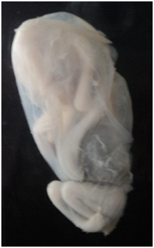
Figure 5 A specimen of a three month old fetus in amniotic sac from embryology section.
Courtesy, Father Muller Medical College Anatomy Museum, Mangalore
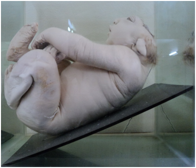
Figure 6 A specimen of fetus with anencephaly. Similar anomalies can be displayed in embryology section.
Courtesy, Yenepoya Medical College Anatomy Museum, Mangalore
Regional gross anatomy: The gross anatomy specimens must be arranged region wise from head and neck and neuroanatomy to thorax and abdomen and the upper and lower extremities.
A cross sectional perspective: Human body can also be studied by sectioning the body at specific intervals. The cross section at a level can be compared with CT-Scan and MRI films taken at the same level. This type of study is useful for medical students. A coronal section of paranasal sinuses is shown in Figure 7. Wooden neuroanatomy models describing cross sections of brainstem at various levels, prepared by the second author are shown in Figures 8‒10. All the nuclei in the model can be removed from the board and the student can be asked to reassemble the nuclei back to its original position and name the nuclei. These models can be applied for objective structured practical evaluation (O.S.P.E).14
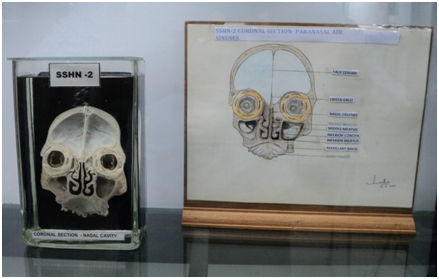
Figure 7 A coronal section of face with the paranasal air sinuses. The specimen is compared to a labeled figure. Similar comparisons can be used for better understanding of sectional anatomy.
Courtesy, Kasturba Medical College Anatomy Museum, Mangalore
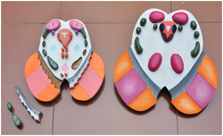
Figure 8 Wooden models of section of midbrain at superior and inferior colliculus.
Courtesy, Yenepoya Medical College Anatomy Museum, Mangalore
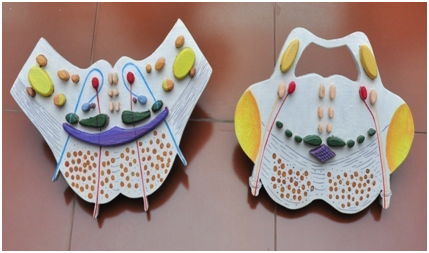
Figure 9 Wooden models of section of pons at facial colliculus and floor of fourth ventricle.
Courtesy, Yenepoya Medical College Anatomy Museum, Mangalore
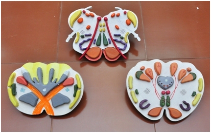
Figure 10 Wooden models of section of medulla at the level of sensory and motor decussation and the olive.
Courtesy, Yenepoya Medical College Anatomy Museum, Mangalore
Note: The models are prepared by the second author. Each nucleus can be removed from the board and the student can be asked to name the nuclei and reassemble the nuclei back to its original position. These models can be applied for objective structured practical evaluation (O.S.P.E) [14].
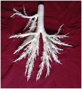
Section with Plastinates: Luminal casts of the tracheo-bronchial tree and whole organ and sheet plastinates can be prepared. A luminal plastinate of tracheobronchial tree of sheep prepared by the first author is shown in Figure 11. It can be compared with the tracheobronchial architecture of humans and other species.15
Imaging and anatomy: Normal and abnormal radiographs can be compared. X-Ray, CT scan and M.R.I must be displayed in this section with relevant literature and audio-visual aids describing the applications of radiology in clinical diagnosis. The section must be designed in a manner that will motivate medical students to learn radiology.
Section for interaction, small group discussion and team based learning with audio-visual aids: It is essential to have a seminar hall, well equipped with the latest audio-visual technology, internet and WI-FI access, multiple access points for computers, a huge sitting arrangement, projectors, speakers and mikes. The hall must be constructed in a manner that will facilitate interaction, small group discussion and team based learning. Electronic screens are used to teach anatomy to medical students in Netherlands, in “The Anatomy Museum of Leiden Medical University”. In Japan, a similar facility with audio-visual aids is used in “The Museum of Kawasaki Medical School”.16 An electronic screen displaying information regarding cleavage, morula, blastocyst, development of germ layers and somites is shown in Figure 12.
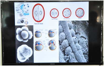
Figure 12 An electronic screen displaying information regarding cleavage, morula, blastocyst, development of germ layers and somites.
Courtesy, Yenepoya Medical College Anatomy Museum, Mangalore
Section with anatomical mannequins: The advances in science have now enabled us to prepare mannequins that can be programmed to practice several skills. Today we have skill labs where mannequins are used for training in ultrasound, laparoscopy, delivery, catheterization and intubation. Anatomical mannequins are also available and can be used in museums as a special attraction. One such mannequin is in the form of virtual table with touch screen and displays anatomy in a three dimensional manner. These mannequins are very expensive and involve huge investment and maintenance expenditure.
Computerized section with multiple access points with internet: A special computerized section can be prepared where data is stored in computers regarding all aspects of museum. The data can be viewed, accessed and transferred into I-pads and pen-drive for viewing at home and is useful for medical students.
Section with three dimensional viewing capabilities: A section with three dimensional viewing capabilities can be created in the museum. Scientific advances have also invented three dimensional holograms which can be used to describe anatomy. These technologies do not need much ground space and can be installed in a very small area. Three dimensional relations of organs can be observed using these advances. In an experiment, colored silicone was introduced into the vessels of the brain. After skilled dissection brain specimens were prepared and 3-D stereoscopic pictures were taken and viewed using a 3-D viewer.17
Mini-theatre: The museum must include a mini-theatre towards the exit, where a brief summary of the history of the museum and the medical college and its future goals can be shown to the public. The medical students depending on the grade must be shown appropriate videos related to gross or clinical anatomy or therapeutic procedures. There must be special simpler videos, designed for school children.
Models: The best models prepared by the faculty can be exhibited in this section. The models of body parts can be exhibited region wise.
Ethnic diversity in anatomy: A section can be prepared that exhibits the ethnic diversity in anatomy. The diversity in external appearance, in the facial skeleton and skull and the diversity in the general skeletal framework can be exhibited in this section. The skeleton and the skulls of different ethnic groups can be compared. The external diversity can be compared using pictures or audio-visual aids.
Feedback section: The last section must be a feedback section that collects information from visitors regarding the museum and their suggestions for improvisation.
Museum staff and guide
The museum staff must include a well trained technician, modeler, artist and adequate number of cleaners based on the area of the museum. There must be guides trained in regional languages to explain anatomy to public. If museum visit is charged then additional staff must be appointed for billing purpose.
The rising concept of digitalization in anatomical pedagogy: A twenty-first century debate
The aim of modern pedagogy is to improve the cognitive and clinical skills of the student. The role of dissection is reduced with more emphasis on imaging and simulation. This form of teaching is known as multimodal teaching and includes small group discussions; demonstrations and team based learning techniques that often use computer and audio-visual aids.18 There are also authors who believe that traditional methods of dissection and demonstration can never be undermined and are equally significant for medical education.19,20 If a museum is scientifically designed as described in the prototype, it can function as an institute where active learning can take place, thus including the museum in the mainstream of medical education. Digitalization of a museum, involves development of anatomy related computer software and audio-visual aids, preferably both in English and in the regional languages, so that both the medical students and the public are benefitted. Anatomy Museum of Leiden Medical University in Netherlands and the Museum of Kawasaki Medical School, in Japan are two such digitalized contemporary museums. These museums use electronic screens and audio-visual aids for educational purpose. The electronic screens display educational information regarding each specimen. In Leiden Medical School, the medical students must visit the museum as a part of their medical training. Faculties from different specialties have prepared audio recordings describing the specimens. These recordings can be downloaded into iPods thus helping the students through the museum. The students are questioned regarding individual specimens following their museum visit.16 Previously, information related to the specimen was stored as pictorial catalogues. The contemporary museums can store all the information as power points, word documents and pictures in computers. Specimen related applied anatomy can also be stored in the form of videos or as internet links to the specific sites. All aspects of anatomy from embryology, histology, gross and applied anatomy concerning the specimen can be stored in specific folders that can be easily accessed and downloaded by the student. Various publications related to the topic can also be stored in separate folders. A computer based pictorial museum catalogue of 150 prosected specimens, has been developed, in the anatomy department of Royal Melbourne Institute of Technology, Australia. The catalogues can also be printed and are designed with the purpose of developing specific teaching modules in the future.21 The latest digital invention that has revolutionized anatomical teaching is the virtual dissection table. The table is designed to create a virtual cadaver having three dimensional viewing ability, virtual knife and touch screen features. The table can be connected to a digital overhead projector and used for anatomy lectures. The virtual table enables better understanding of radiological and cross sectional anatomy in a three dimensional manner. The three dimensional holograms will also soon find a place in anatomical teaching. The advances in science and technology may over years replace cadaveric dissection, with digital dissection using a virtual cadaver. However, the topic of cadaveric dissection verses digitalization continues to be an anatomical debate of the twenty-first century.
Museum maintenance
Good maintenance ensures that the museum remains in good shape for long periods of time. Museum maintenance involves the following steps:-
Prototype of a digitalized anatomy museum
A prototype of a digitalized anatomy museum designed by the authors is shown in Figure 13.
The anatomy museums have evolved significantly over the centuries from an institute housing models, to formalin preserved specimens to palatinates. It is essential to constantly upgrade the methods of display in anatomy museums. The future generations live in world that is completely digitalized. Hence, all the modern inventions such as computer, internet, Wi Fi, electronic screens, projectors, 3-D viewing capabilities, 3-D holograms and anatomical mannequins must be used to teach anatomy in anatomy museums. It is also essential to incorporate, a visit to anatomy museums as a part of regular curriculum, in schools and in undergraduate medical education. An anatomy museum is not meant only for display of specimens. It has to be designed, to function as an institute where active learning can take place, thus including the museum in the mainstream of medical education. The future of anatomy will certainly be more and more digitalized. However, the traditional techniques of model making, dissection and plastination will always have a special place in all anatomy museums for time immemorial.
The authors are grateful to the authorities of all the medical colleges for providing the permission to study the museums and for allowing photography. A special thanks to the authorities of Kasturba Medical College, Manipal where the first author completed his postgraduate thesis in anatomy museums.
Author declares that there is no conflict of interest.

©2016 Kamath, et al. This is an open access article distributed under the terms of the, which permits unrestricted use, distribution, and build upon your work non-commercially.