Journal of
eISSN: 2373-4426


Case Report Volume 8 Issue 3
1Senior specialist pediatric, Pediatric Department Ibri Hospital, Sultanate of Oman
2Medical Officer, Radiology Department Ibri Hospital, Sultanate of Oman
3Pediatric radiologist Specialist, Radiology Department Ibri Hospital, Sultanate of Oman
Correspondence: Farida Ambusaidi, Pediatric radiologist Specialist, Radiology Department Ibri Hospital, Sultanate of Oman, Tel 96899793810
Received: March 28, 2018 | Published: June 28, 2018
Citation: Al-Majrafi AS, Kalbani NKA, Ambusaidi F. Rare congenital fistula connection between right subclavian artery and superior vena cava presenting in neonate with congestive cardiac failure. J Pediatr Neonatal Care. 2018;8(3):148-149. DOI: 10.15406/jpnc.2018.08.00328
This case report describes a congestive cardiac failure in neonate secondary to rare congenital arteriovenous fistula between the right subclavian artery and superior vena cava. The abnormality was initially discovered by echocardiography and then confirmed by CT angiography. The neonate got successful surgical repair and uneventful post surgical period. Intra-thoracic congenital arteriovenous malformations are very rare and presenting early with cardiac failure, which is challenging for diagnosis and treatment. However early identification and treatment is the most influencing factor in mortality and morbidity of these cases.
Keywords: arteriovenous malformation, subclavian artery, fistula, congestive cardiac failure
Intra-thoracic arteriovenous malformations are uncommon. As the most reported localizations of AVM are the head (vein of Galen malformation), the abdomen (infantile hepatic hemangioma), the neck and extremities.1 Arterio venous malformation divided into congenital and acquired. The congenital forms are even more uncommon and patients present with variable symptoms that make the diagnosis more challenging.2,3 A congenital Aortocaval fistula from subclavian artery to the superior vena cava (SVC) may represent a subclass of this condition.
Our patient is full term to primigravida mother born by spontaneous vaginal delivery with good Apgar score and birth weight of 3.48 kg. Baby required no resuscitation. No risk factors for sepsis. Baby developed tachypnea at 2 hours of life, so shifted to Special care Baby Unit (SCBU). Examination revealed bruit over right subclavian area.
Echocardiography was done on day one of life and showed:
Impression: Extra cardiac arteriovenous shunt, Right subclavian artery (SCA) to SVC
Baby started on anti failure medications. The baby deteriorated on day 2 of life, so incubated. Echo repeated with same findings plus newly developed pulmonary hypertension (Figure 1) (Figure 2).

Figure 1 Short axis view Echocardiography for the AV connection between the dilated SCA to Dilated SVC.
Baby started on anti failure medications. The baby deteriorated on day 2 of life, so incubated. Echo repeated with same findings plus newly developed pulmonary hypertension.
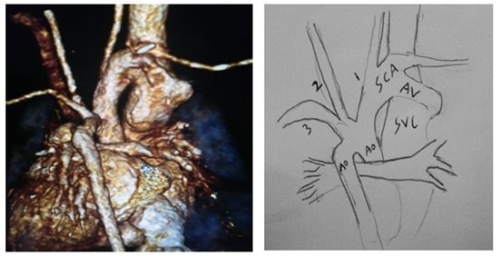
*CT Angiography showed:

Figure 2A, Figure 2B 3D CT angio for the great arteries and the AV malformation, (B) Diagram illustration of the AV malformation in figure (A).
*USG head was done and showed no obvious vein of Galen malformation.
The baby was transferred to the national cardiac center where he got repair of right subclavian artery to superior vena cava arterio-venous fistula (ligation and division) and ligation of patent ductus arteriosus.
Aortocaval fistulas are rare cause of left-to-right-shunt resulting in neonatal congestive heart failure. The vessels initially develop from the mesenchyma in an undifferentiated form till give fully developed veins and arteries. So, they develop from same primordial network of vessels. Congenital arteriovenous fistulas may be the result, if these early connections of both arteries and veins persist. Hereby congenital AVM is a mal-development of blood vessels, with preservation of one or more primitive direct communications between arterial and venous channels.4,5 Histological, the fistula connection shows hyperplasia of elastic fibers of the arterialized vein, ranging from 41 to 82% .6,7 There are few cases in literature similar to our case, right subclavian artery to SVC fistula reported in 1977 by Arkell and Lawson8, in 2011 case by Balakrishnan et al.,9 and another case by Awasthy et al.10. Three case reported by Gutierrez et al.,2 two of them showed a fistula between left subclavian artery-to-innominate vein and draining into markedly dilated superior vena cava. Sapire et al.11 In 1983 reported a case of arterio-venous fistula between the subclavian artery and in nominate vein presented with heart failure in neonatal period.11 In 2000 Dogan et al.,12 & Tatum et al.,13 in 2006 reported congestive heart failure due to subclavian artery to subclavian vein fistula. In 2016, Sami Jabari & Robert Cesnjevar14 reported a case of fistulatous connection between Brachiocephalic trunk and superior vena cava. The echo cardio graphy is simple fast screen tools for cardiac abnormality and first alert to such entity. The confirmation by using 64 slice CT scan was hand-to-hand a great support that confirm the diagnosis which facilitate the transfer of patient to the appropriate institution to get the proper treatment.
The early presentation of heart failure in neonatal period necessitate extensive work up excluding all arteriovenous malformation in heart, head, chest and abdomen. This case highlights a rare congenital arteriovenous fistula connection between subclavian artery and superior vena cava. Echocardiography is important non invasive tool in diagnosing and raising suspicion of congenital Aortocaval arteriovenous fistula. CT angiography was complementary tool to diagnose the malformation.
The author declares there is no conflict of interest.

©2018 Al-Majrafi, et al. This is an open access article distributed under the terms of the, which permits unrestricted use, distribution, and build upon your work non-commercially.