
Case Report Volume 2 Issue 2
Pyloric Duplication: About Two Cases
Y Ben Ahmed,
Regret for the inconvenience: we are taking measures to prevent fraudulent form submissions by extractors and page crawlers. Please type the correct Captcha word to see email ID.

S Ghorbel, A Charieg, F Nouira, R Khemekhem, S Jlidi, B Chaouchi
Department of Pediatric surgery, Tunis Medical School, Tunisia
Correspondence: Y Ben Ahmed, Pediatric surgeon, Department of Pediatric surgery, Tunis Medical School, Tunis, Tunisia, Tel 33624902112
Received: October 31, 2014 | Published: April 20, 2015
Citation: Ahmed YB, Ghorbel S, Charieg A, Nouira F, Khemekhem R, et al. (2015) Pyloric Duplication: About Two Cases. J Pediatr Neonatal Care 2(2): 00065. DOI: 10.15406/jpnc.2015.02.00065
Download PDF
Abstract
Pyloric duplication cyst is a rare congenital anomaly. Few cases have been described in the literature. We report two cases of 2 and 5year-old girl. The diagnosis was suspected on ultra sonography and upper gastrointestinal series and confirmed by surgery and histopathologic examination. Resection was total in one case and partial in the other. Pyloroplasty was associated. Post operative was unfaithful.
Keywords: duplication, pylorus, resection, pyloroplasty
Introduction
Duplication cysts of the pylorus are the rarest of gastrointestinal duplications. Few cases have been described in the literature.1 In most cases preoperative diagnosis is not made.2 To our knowledge, no more than 20 cases of pyloric duplication have been reported in the English literature,1−18 most of them were extra luminal masses. Here in, we describe two cases with pyloric duplication cyst.
Case 1
A five year old girl was hospitalized because she has suffered from abdominal pain and non bilious vomiting for a month. Her physical examination was unremarkable except epigastric tenderness. Biologic exams were normal specifically amylasemia. An upper gastrointestinal barium contrast study revealed in spite of a sus carina reflux, a pyloric and antrum deformation, with an extrinseque compression. Oesogastric fibroscopy showed a deformation of pylorus. During surgery, pyloric cystic duplication of 2cm*3cm was noticed (Figure 1). By opening the mass, we found 3 cystic formations; the bigger one has a common wall with the posterior face of the pylorus and the antrum with a narrowed communication. The duplication was resected as well as the common wall and a pyloroplasty was performed (Figure 2). There were no postoperative problems and the post operative barium - meal examination was normal.
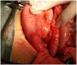
Figure 1 A pyloric cystic duplication of 2 cm*3cm.
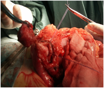
Figure 2 Duplication resection as well as the common wall.
Case 2
A two year old girl was presented in emergency for paroxystic abdominal pain without vomiting or other signs. The physical exam showed an epigastrium mass of 3cm which is round, tense, smooth and non tender beneath the umbilicus. Biologic exams were normal. Abdominal ultrasound with computer tomography revealed a cystic epigastric mass of 4.3cm with a wall that took contrast (Figure 3). Around this mass there was an ultrasound aspect of volvulus (Figure 4). A barium-meal examination demonstrated delayed gastric emptying through a narrowed pyloric antrum, which was distorted by a non communicating mass (Figure 5). At laparotomy, a cystic mass measuring approximately 4 × 3cm was identified anterior and lateral to the pyloric channel, sharing a common wall. The mass compressed the pyloric channel, resulting in partial gastric outlet obstruction. The duplication cyst did not communicate with the pyloric channel. Most of the cyst was excised, and the mucosa of the remnant cyst wall was cauterized. Histologic examination revealed gastric mucosa with a smooth muscle coat, which was consistent with a pyloric duplication cyst. No aberrant tissue was seen. The postoperative course was uneventful. The patient was asymptomatic 4years later.
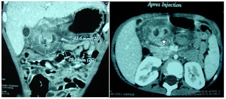
Figure 3 Computer tomography: Cystic epigastric mass with a fine wall ( ☆ ) that took contrast.
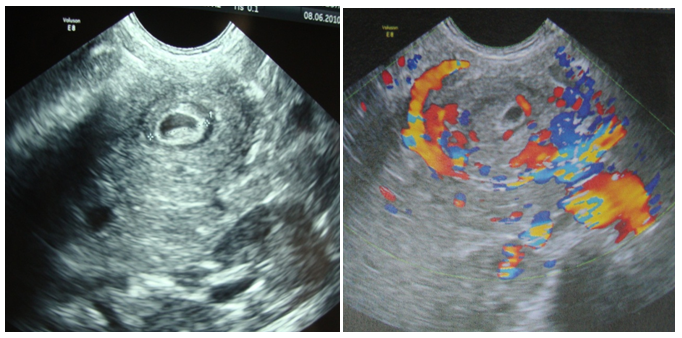
Figure 4 Ultrasound aspect of volvulus around the mass.
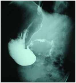
Figure 5 A barium-meal examination: Narrowed pyloric antrum distorted from below by a non communicating mass.
Discussion
- Congenital duplications are uncommon congenital lesions. They may arise anywhere in the gastrointestinal tract from the mouth to the anus with an incidence of 1 in 4500 births.9,13 This spherical or tubular cystic structure is lined with alimentary tract epithelium and has smooth muscle in its wall. Less than 4% of all gastrointestinal duplications (GID) involve the stomach.3,19 Pyloric duplication cysts are uncommon lesions; most of them are extraluminal masses. The female-to-male ratio is 2:1. The vast majority is discovered in the 1styear of life. Only one third of the duplications is present in the neonatal period.19,20 In literature, ages vary between 1 day and 2years old.15,21 Our patients are the oldest reported in case of pyloric duplication.
- Gastrointestinal duplication (GID) has an embryonic origin, it probably develops in the sixth week of embryonic life but the etiology has not been elucidated. Several theories have been proposed to explain their development. These include the persistence of embryonic diverticulae during alimentary development, abortive attempts at twinning, intrauterine vascular accidents, and recanalization with fusion of embryological longitudinal folds.2,3,11,20,22 However, none of these theories alone is able to explain the full diversity of these lesions. The most attractive theory is the split notochord syndrome (adhesions of entoderm to neuroectoderm).
- The presentation of gastrointestinal duplication can vary according to the age and the localization. Some GID may be totally asymptomatic, identified on routine physical examination or during investigations for other problems. Common clinical manifestations of pyloric duplications include a palpable abdominal mass and non bilious vomiting which is almost present in all the cases of literature.1−18 Bleeding, intussusception can be an occasion for discovering such unusual anomalies.3,20 Most pyloric duplication cysts have no communication with the adjacent bowel tract and have no complications.8 In the first case pyloric duplication have communication with digestive tract.
- With the increasing use of prenatal ultrasound scan, some are now being identified In utero.23 Five cases were reported in prenatally diagnosis.19 The accurate preoperative diagnosis of GID, especially pyloric duplication, is challenging. Conventional plain radiographic studies are nonspecific. Upper gastrointestinal tract contrast studies may show an extrinsic filling defect. Ultrasound is the most common modality. It may reveal the inner hyperechoic and outer hypoechoic layers (double-wall sign) that are typical of duplication cysts considered by Deftereos as a confirmation of the diagnosis.4,24 However, in the case we described, ultrasound showed, only, an anechoic cystic mass with a single hyperechoic wall, and the typical double-wall sign was not reached. Computer tomography CT is not only useful for making a correct diagnosis of the cysts duplications but also provides better information than US regarding the exact extension and limits of cystic masses.8 Nuclear scans using radioisotopes may show tracer uptake by ectopic gastric mucosa. Magnetic resonance imaging studies are useful in demonstrating the relationship with other structures.4,15 If, after appropriate investigations, the diagnosis remains unclear, we suggest that laparoscopy should be used to confirm the diagnosis.22 Hypertrophic pyloric stenosis, pyloric atresia and congenital antral webs should be included in the differential diagnosis in case of newborn. We can also include mesenteric cysts, retroperitoneal cysts, lymphangioma, and in female patients, ovarian cysts.10
- Definitive treatment of GID involves ideally total excision, which often requires resection of the adjacent bowel as duplication cysts usually share a common wall with the bowel and so a restoration of continuity of the lumen.25 However, in some cases, because of extensive size or location, alternate procedures such as subtotal excision with removal of mucosa or its cauterization without resection of the contiguous pylorus can be made. In our second case, considering important adhesions, we did subtotal excision. This approach allows rapid postoperative recovery, but any procedure should ideally ensure removal of mucosa to prevent subsequent malignant transformation into carcinoma in later adulthood.1,2 Laparoscopy should be utilized liberally in patients with atypical presentation of inflammatory bowel disease or when the diagnosis of abdominal cystic masses in neonates or infants is unclear.18,25
Conclusion
Duplications involving the pylorus are particularly rare. Diagnostic studies, including ultrasonography, an upper gastrointestinal barium study, and regarding the case endoscopy, and CT, are useful in establishing the preoperative diagnosis. Surgery is the undisputed mainstay of management. It ranges from simple excision to pyloroantrectomy. In all surgical procedures, it is essential to remove the entire lining of the duplication cyst.
Acknowledgments
Conflicts of interest
Author declares there are no conflicts of interest.
Funding
References
- Tang XB, Bai YZ, Wang WL. An intraluminal pyloric duplication cyst in an infant. J Pediatr Surg. 2008;43(12):2305−2307.
- Mărginean CO, Mărginean C, Horváth E, et al. Antenatally diagnosed congenital pyloric duplication associated with intraluminal pyloric cyst - rare entity case report and review of the literature. Rom J Morphol Embryol. 2014;55(3):983−988.
- Saad DF, Gow KW, Shehata B, et al. Pyloric duplication in a term newborn. J Pediatr Surg . 2005;40(7):1209−1210.
- Chin AC, Radhakrishnan RS, Lloyd J, et al. Pyloric duplication with communication to the pancreas in a neonate simulating hypertrophic pyloric stenosis. J Pediatr Surg . 2011;46(7):1442−1444.
- Iwasaki M, Nishimura A, Kamimura R, et al. Pyloric duplication cyst in an infant. Pediatr Int. 2009;51(1):146−149.
- Novak RW, Boeckman CR. Pyloric duplication presenting with hemorrhagic ascites. Clin Pediatr (Phila) . 1983;22(5):386−388.
- Keramidas DC, Doulas NL, Anagnostou D. Duplication of the pylorus: an elusive diagnosis. Z Kinderchir Grenzgeb. 1980;29(4):370−374.
- Grosfeld JL, Boles ET Jr, Reiner C. Duplication of pylorus in the newborn: a rare cause of gastric outlet obstruction. J Pediatr Surg. 1970;5(3):365−369.
- Langer JC, Superina RA, Payton D. Pyloric duplication presenting with gastric outlet obstruction in the newborn period. Pediatric Surgery International. 1988;4(1):63−65.
- Mogilner JG, Vinograd I, Presman A, et al. Pyloric duplication in children. Pediatric Surgery International . 1990;5(1):61−63.
- Shah A, More B, Buick R. Pyloric duplication in a neonate: a rare entity. Pediatr Surg Int. 2005;21(3):220−222.
- Sinha CK, Nour S, Fisher R. Pyloric duplication in the newborn: a rare cause of gastric outlet obstruction. J Indian Assoc Pediatr Surg. 2007;12(1):34−35.
- Patel MP, Meisheri IV, Waingankar VS, et al. Duplication cyst of the pylorus - a rare cause of gastric outlet obstruction in the newborn. J Postgrad Med. 1997;43(2):43−45.
- Upadhyaya VD, Srivastava PK, Jaiman R, et al. Duplication cyst of pyloroduodenal canal: a rare cause of neonatal gastric outlet obstruction: a case report. Cases J. 2009;2(1):42.
- Trainavicius K, Gurskas P, Povilavicius J. Duplication cyst of the pylorus: a case report. J Med Case Rep. 2013;7:175.
- Mirza B. Pyloroduodenal duplication cyst: the rarest alimentary tract duplication. APSP J Case Rep. 2012;3(3):19.
- Sharma D, Bharany RP, Mapshekhar RV. Duplication cyst of pyloric canal: a rare cause of pediatric gastric outlet obstruction: rare case report. Indian J Surg. 2013; 75(Suppl 1):322−325.
- Bommen M, Singh MP. Pyloric duplication in a preterm neonate. J Pediatr Surg. 1984;19(2):158−159.
- Murty TV, Bhargava RK, Rakas FS . Gastroduodenal duplications. J Pediatr Surg. 1992;27(4):515−517.
- Hamada Y, Inoue K, Hioki K. Pyloroduodenal duplication cyst: case report. Pediatr Surg Int . 1997;12(2-3):194−195.
- Zhou LQ, Wang BH, Zuo YH. A case report of inborn pyloric duplication. Zhongguo Dang Dai Er Ke Za Zhi . 2007;9(5):421.
- Puligandla PS, Nguyen LT, St-Vil D, et al. Gastrointestinal duplications. J Pediatr Surg. 2003;38(5):740−744.
- Deftereos S, Tsalkidis A, Soultanidis H, et al. Pyloric duplication: ultrasound appearance. Minerva Pediatr. 2006;58(4):395−397.
- Goyert GL, Blitz D, Gibson P, et al. Prenatal diagnosis of duplication cyst of the pylorus. Prenat Diagn. 1991;11(7):483−486.
- Cooper S, Abrams RS, Carbaugh RA. Pyloric duplications: review and case study. Am Surg. 1995; 61(12):1092−1094.

©2015 Ahmed, et al. This is an open access article distributed under the terms of the,
which
permits unrestricted use, distribution, and build upon your work non-commercially.


 International Childhood Cancer Day is observed on 15 February 2026 to raise awareness about childhood cancers and to highlight the medical, developmental, and supportive care needs of affected children and their families. This day emphasizes the importance of early diagnosis, pediatric care, and continued research to improve survival and quality of life in children with cancer.
Researchers and healthcare professionals are encouraged to submit their original research articles, reviews, and clinical studies related to pediatric oncology, neonatal care, and child health. Manuscripts submitted on the occasion of International Childhood Cancer Day will be eligible for a special publication discount of 30–40% in the Journal of Pediatrics & Neonatal Care (JPNC).
.