Proceeding Volume 5 Issue 6
Pulmonary Air Leak in a Newborn
Khawaja Mahmood
Regret for the inconvenience: we are taking measures to prevent fraudulent form submissions by extractors and page crawlers. Please type the correct Captcha word to see email ID.

Medical College of Georgia, Georgia
Correspondence: Khawaja Mahmood MD FAAP, Associate Professor of Pediatrics, Medical College of Georgia, Georgia
Received: November 15, 2016 | Published: November 29, 2016
Citation: Mahmood K (2016) Pulmonary Air Leak in a Newborn. J Pediatr Neonatal Care 5(6): 00206. DOI: 10.15406/jpnc.2016.05.00206
Download PDF
Pulmonary air leak
- Pulmonary air leak occurs more frequently in the newborn period than at any other time of life.
- Air escapes the lungs into extra-alveolar spaces-resulting disorder depends on the location of air.
- Most common conditions.
- Pneumothorax, pneumomediastinum, and pneumopericardium.
- Rarer conditions.
- Subcutaneous emphysema and pneumoperitoneum
Risk factors
- Most cases of air leaks occur in newborns with underlying lung disease.
- Preterm infants are at increased risk because they frequently have Respiratory Distress Syndrome (RDS).
Pneumothorax
- Air in the space between the parietal and visceral pleura.
- Usually show signs of respiratory distress such as tachypnea, grunting, pallor, and cyanosis.
- Findings on Physical exam.
- Chest Asymmetry.
- Decreased breath sounds on affected side.
- Shift of the PMI away from the affected side.
Diagnosis of a pneumothorax
- Should be suspected in any newborn with the sudden onset of respiratory distress.
- Transillumination of the chest may help make the diagnosis:
- A pnuemothorax lights up the affected hemithorax.
- AP Chest radiograph:
- Air in the pleural space.
- Flattening of the diaphragm on the affected side.
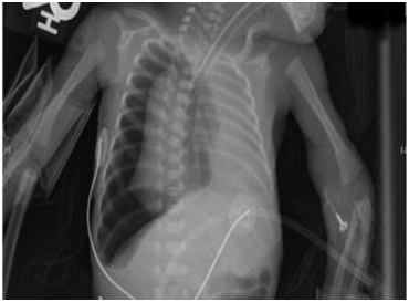
- Shift of the mediastinum away from the pnuemothorax (Figure 1).

Figure 1 Neonate with Right Tension Pneumothorax.
Management of a pneumothorax
- Infants without a continuous air leak or respiratory distress can be closely monitored.
- Oxygen supplementation.
- Thoracentesis for emergent treatment of a symptomatic pneumothorax.
- Chest tube placement for definitive drainage.
Congenital lobar emphysema
- Developmental anomaly of the lower respiratory tract that is characterized by hyperinflation of one or more of the pulmonary lobes.
- Rare with a prevalence of 1 in 20,000 to 1 in 30,000.
- Males affected more than females in a 3:1 ratio.
- 50% of cases occur in the first 4 weeks after birth.
- 75% of cases are found in infants < 6 months of age.
Congenital lobar emphysema
Characterized by:
- Difficulty in breathing or very rapid respiration in infancy.
- Enlarged chest due to over inflation of at least one lobe of the lung.
- Compressed normal lung tissue in the section of the lung closest to the diseased lobe.
- Bluish color of the skin (cyanosis.)
- Underdevelopment of the cartilage that supports the bronchial tube (bronchial hypoplasia).
Diagnosis of congenital lobar emphysema
- Chest X-Ray, CT, MRI can determine which part of the lung, which lobe is affected, and to what degree.
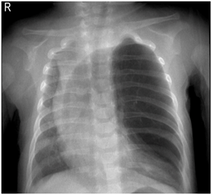
- Lung function tests are also helpful studies to determine which part of the lung are affected and if surgery is necessary (Figure 2).

Figure 2 Neonate with Congenital Lobar Emphysema.
Management of congenital lobar emphysema
- Depends on the extent of the damage to the lungs at the time of diagnosis.
- If lung damage is limited, disease may not cause any adverse affects-closely monitor.
- If condition affects the patient’s ability to breath-Surgical removal (resection) of the affected lobe of the lung or the whole lung on the affected side.
Acknowledgments
Conflicts of interest
Author declares that there is no conflict of interest.
References

©2016 Mahmood. This is an open access article distributed under the terms of the,
which
permits unrestricted use, distribution, and build upon your work non-commercially.


