Journal of
eISSN: 2373-4426


Case Report Volume 11 Issue 1
1Senior Specialist, Department of Neonatology, Latifa Women and Children Hospital, UAE
2Consultant, Department of Neonatology, Latifa Women and Children Hospital, UAE
3Consultant and Head, Department of Neonatology, Latifa Women and Children Hospital, UAE
Correspondence: Tushar Kulkarni, Senior Specialist Neonatology, Neonatal Intensive Care Unit, Latifa Women and Children Hospital, Oud Metha Road, Dubai, United Arab Emirates
Received: February 21, 2021 | Published: March 27, 2021
Citation: Kulkarni T, El-Atawi K, Elhalik M. Neonatal Tuberous Sclerosis: is skin really the window? J Pediatr Neonatal Care. 2021;11(1):23-25. DOI: 10.15406/jpnc.2021.11.00437
We report a case of a neonate who was diagnosed to have intracardiac mass as a fetus and presented with white linear papules that was diagnosed to be Linear Verrucous Epidermal Nevus. Apart from the intracardiac mass, most likely rhabdomyoma, MRI Brain also showed presence of tubers and the Next Generation Sequence Analysis confirmed the diagnosis of Neonatal Tuberous Sclerosis. The neonate remained asymptomatic and was discharged home and remains under close follow up without any symptoms. It is thus suggested that Linear Verrucous Epidermal Nevus, a cutaneous manifestation not described with Tuberous Sclerosis could be considered under the ever-expanding cutaneous signs of Tuberous Sclerosis and should alert the physician toward its possibility.
Keywords: neonate, tuberous sclerosis, unusual cutaneous manifestation, linear epidermal verrucous nevus
LVEN, linear verrucous epidermal nevus; TSC, tuberous sclerosis complex; MRI, magnetic resonance imaging; LVOT, left ventricular outflow tract; NICU, neonatal intensive care unit; PDA, patent ductus arteriosus; FLAIR, fluid attenuated inversion recovery; EEG, electroencephalogram; NGS, next generation sequencing; mTOR, mammalian target of rapamycin; FGF3R, fibroblastic growth factor 3 receptor
Tuberous Sclerosis is a neurocutaneous disorder with an autosomal dominant pattern of inheritance and is rarely seen in the neonatal age group. Among the myriad of cutaneous manifestations, of which most present later in the childhood, only ‘ash leaf’ macules are known to present in the neonates. It is important to evaluate neonates presenting with isolated typical and atypical cutaneous features as described below for probable Tuberous Sclerosis to aid early diagnosis and management planning for associated multisystem manifestations that can present later and can be debilitating. Our case report highlights the importance of being aware of the atypical cutaneous manifestations of Tuberous Sclerosis in the neonatal age group.
Following a relatively uncomplicated pregnancy a 35-year-old para 4 lady, non-consanguineously married, with previous 2 healthy boys and 2 healthy girls delivered a girl after term pregnancy. Her antenatal scan at 28 weeks gestation detected single intracardiac mass in the fetus for the first time. A fetal echocardiography done at 35 weeks of gestation showed multiple echogenic masses of variable size in both the cardiac ventricles with the largest one at the interventricular septum pointing toward the left ventricular outflow tract (LVOT) without causing any functional obstruction. The pregnancy was monitored with regular scans and the masses remained the same and did not cause any hemodynamic compromise. Due to the possibility of Tuberous Sclerosis, this couple was screened for any cutaneous manifestations and none were found. They were appropriately counseled by the multidisciplinary team including FetoMaternal Medicine specialist, Neonatologist, and the Pediatric Cardiologist especially for the strong possibility of fetus having Tuberous Sclerosis. Following delivery, the girl was noted to weigh 3170 grams (44th centile), have a head circumference of 34cms (54th centile) and length of 49 cms (46th centile). She was admitted to the NICU for further work up and observation of her hemodynamic status related to the intracardiac masses.
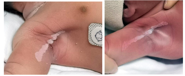
She remained hemodynamically stable during her entire NICU stay. Postnatal Echocardiography showed 3 intracardiac masses, all three arising from interventricular septum with the biggest measuring 20x17x8mm pointing toward subaortic region and partly encroaching over the LVOT. The other two small masses were both in the right ventricle pointing toward the apex. Additionally, a tiny PDA (Patent Ductus Arteriosus) was observed. On further evaluation, the baby was noted to have a macerated linear verrucous lesion along the Blaschko’s lines in the right axillary region measuring 8cms in length– ‘Linear Epidermal Verrucous Nevus (LVEN)’ (Figure 1). The maceration improved and left a pinkish background over the next few days. On further observation, few faint hypopigmented macules (4-5 each measuring nearly 6-7 mm) were also noted on the back. Neuroimaging revealed presence of multiple T1 hyperintense and T2 (FLAIR) hypointense subependymal hamartomas and linear /nodular white matter hyperintensities on either side- most likely cortical and subcortical tubers along with white matter hyperintensities in the region of centrum semiovale showing radiating pattern, all suggesting cortical dysplasia.1,2 EEG was normal. Further imaging of kidneys and evaluation of retina did not reveal any abnormality. Presence of more than 2 major criteria (intracardiac masses- most likely Rhabdomyoma, cortical dysplasia, hypomelanotic- ‘ash leaf’ macules) almost certainly pointed towards Tuberous Sclerosis (Table 1).1 Genetic Analysis done with Next Generation Sequencing (NGS) revealed pathogenic mutation of TSC 1 (Tuberous Sclerosis Complex1) gene. Familial Mutation Screening did not reveal similar findings in the parents, pointing towards de novo causation of Tuberous Sclerosis in the baby. This baby girl remained hemodynamic stable, had no seizures, and could be discharged home after 7 days of hospital stay with a multidisciplinary follow up (Neonatology, Pediatric Cardiology, Pediatric Neurology, Pediatric Nephrology, Dermatology and Pediatric Ophthalmology). Her weight was 3100g on discharge and the skin lesions namely, LVEN had become fainter and the ‘ash leaf spots’ persisted. She remains asymptomatic during follow up appointments.

Figure 1 Image of LVEN- Linear Verrucous Epidermal Nevus at birth (on left) and after 2-3 days (on right).
|
No. |
Major Criteria |
|
Minor Criteria |
|
1 |
Hypomelanotic macules (>3, >5mm in diameter) |
1 |
‘Confetti’ skin lesions |
|
2 |
Angiofibromas (>3) or fibrous cephalic plaque |
2 |
Dental enamel pits (> 3) |
|
3 |
Ungual fibromas (>2) |
3 |
Intraoral fibroma (>2) |
|
4 |
Shagreen patch |
4 |
Retinal achromic patch |
|
5 |
Multiple retinal hamartomas |
5 |
Multiple renal cysts |
|
6 |
Cortical dysplasia |
6 |
Nonrenal hamartomas |
|
7 |
Subependymal nodules |
|
|
|
8 |
Subependymal giant cell astrocytoma |
|
|
|
9 |
Cardiac rhabdomyoma |
|
|
|
10 |
Lymphangioleiomyomatosis |
|
|
|
11 |
Angiomyolipomas (>2) |
|
|
Table 1 Clinical Criteria for the diagnosis of Tuberous Sclerosis Complex (TSC)
Definitive Diagnosis of TSC: Presence of Pathological mutation of TSC1/TSC2 gene OR presence of 2 major criteria OR presence of 1 major + >2 minor criteria
Probable Diagnosis of TSC: Presence of 1 Major OR >2 minor criteria
(Adapted from Portocarrero LKL. An Bras Dermatol. 2018;93(3):3233, Krueger DA. Pediatr Neurol. 2013; 49:2552)
TSC, an autosomal dominant condition, is classified as a neurocutaneous disorder associated with hamartomas in skin, central nervous, heart, lungs and kidneys with variable phenotypic appearance making its recognition difficult, especially in the earlier childhood. Incidence of 1:6000 to 1:10,000 among live births has been reported.1-3 Revised Diagnostic Criteria (Table 1)2,3 published in 2012 by International Tuberous Sclerosis Complex Consensus Group1 provide evidence-based recommendations that are simple, easy and reliable. Using genetic mutational analysis as an independent criterion and classifying cases as probably affected and possibly affected is the most striking change from previous criteria.1,4,5
Based on the same criteria, our case already had >2 major (cardiac rhabdomyoma + cortical dysplasia + ‘ash leaf spots’). Later, mutational analysis of TSC 1 gene confirmed the already certain diagnosis. Davis et al.,6 in one of the largest prospective studies among infants with TSC, reported hypomelanotic macules (94%) as one of the commonest diagnostic features of TSC that are not only the earliest but are the ‘only’ cutaneous manifestation of neonatal TSC.7
Although our case did demonstrate ‘ash leaf spots’- which is a clear criteria for diagnosis of Neonatal Tuberous sclerosis, the presence of linear verrucous epidermal nevus (LVEN), that appeared macerated initially at birth and then later on cleared, was perplexing. Epidermal nevi are benign, hamartomal growths of skin, characteristically along the lines Blaschko and may be present at birth but generally are not hereditary, arising only due to somatic mutations. LVEN may be isolated or be a part of epidermal nevus syndrome. Differential diagnoses of LVEN also include linear planus, psoriasis, lichen striatus and porokeratosis.8 In our case, biopsy of the lesion was not considered as the diagnosis of Tuberous sclerosis was certain.5 A case described by Elam et al.9 showed similar white epidermal nevus in a neonate on day 1 of life but these appeared as plaques and papules that later on was diagnosed as TSC. Their case presented with only nevi at birth but later developed hypopigmented macules followed by seizures at age 3 months prompting the diagnosis of Tuberous Sclerosis. Co-existence of LVEN and Tuberous Sclerosis simultaneously can be explained by a common regulatory pathway.3 It is reported that, 42% of epidermal nevi are actually due to mutations that produce proteins which act as upstream regulators of the TSC1 and TSC2 genes.9-11 Similarities between Linear Sebaceous Nevus Syndrome, another kind of hamartoses and Tuberous Sclerosis have been described in the past12 and recently, newer osseocutaneous manifestations with TSC have also been described.13
Progress of LVEN from being macerated to pinkish color can be explained by the initial maceration due to amniotic fluid hydration in utero followed by quick keratinization.14 In our case, despite LVEN, presence of other classic features related to TSC prompted the diagnosis. We understand that the diagnosis would have been more difficult had it been if LVEN was an isolated skin finding. Working up for probable TSC based on isolated findings can cause unnecessary anxiety to the family and financial costs of screening that generally have a low diagnostic yield. Moreover, misattribution of such skin lesions to a potentially life modifying diagnosis such as TSC would certainly not be worthwhile. Need of the hour would be to identify features that increase suspicion of TSC and decide where extensive TSC-related work-up may or may not be required. An algorithm that evaluates individuals presenting with one skin finding suggestive of TSC has been described.15 Based on clinical diagnostic workup, remaining patients should receive a diagnosis of probable TSC1-3 and the parents be informed that the likelihood of disease onset and suspicion.15,16 At the same time, it is also important to note that the cutaneous manifestations of TSC are ever expanding and care givers should be aware of some atypical skin signs that could prompt possibility of TSC.
In our case, although initially thought to be coincidental finding, it can be purported that LVEN could thus, be considered a manifestation of Tuberous sclerosis, acting as a window to the inner pathology making it even more important for pediatricians and parents to be aware of seizure activity in neonates who present with multiple scattered epidermal nevi that are not typical of epidermal nevus syndrome variants.9
Description of well-established diagnostic criteria has helped pediatricians promptly diagnose, refer, manage and undertake surveillance of children with TSC. Apart from a constellation of typical TSC-associated cutaneous manifestations including the classical ‘ash leaf spots’ in the neonate, atypical skin lesions such as LVEN can also develop at birth or during early childhood, highlighting the role of awareness among the pediatricians of TSC-associated typical and atypical skin lesions.
Nil.
None.

©2021 Kulkarni, et al. This is an open access article distributed under the terms of the, which permits unrestricted use, distribution, and build upon your work non-commercially.