Journal of
eISSN: 2373-4426


Case Report Volume 12 Issue 2
1Professor of Pediatrics Neonatology and Pediatric Intensive Care Balamand University, Senior Lecturer at Saint George University Hospital, Head of Pediatric Department, Beirut Quarantina Governmental Hospital, Tresurer UMEMPS, Lebanon
2Vascular surgeon and vascular intervention, Saint George Universty Hospital, Lebanon
3Saint George University Hospital and Balamand University, Lebanon
Correspondence: Robert Sacy, Professor of Pediatrics Neonatology and Pediatric Intensive Care Balamand University, Senior Lecturer at Saint George University Hospital, Head of Pediatric Department, Beirut Quarantina Governmental Hospital, Tresurer UMEMPS, Lebanon
Received: March 15, 2022 | Published: April 11, 2022
Citation: Sacy R, Chamseddine A, El Masri MPGY. Cystic Hygroma: 3 case reports of head and neck cystic hygroma in neonates. J Pediatr Neonatal Care. 2022;12(2):58-59. DOI: 10.15406/jpnc.2022.12.00455
Objective: Congenital malformations of the lymphatic system, known as lymphatic lymphangiomas, are benign vascular tumors that are usually detected on prenatal ultrasound, and most commonly occur in the head and neck region. The aim of this paper it to report our experience with three cases of cystic hygromas along with a review of the literature.
Materials and methods: Herein we present three cases of cystic hygroma detected within the neonatal period treated with ultrasound guided aspiration.
Results: Following the procedure, two babies had recurrence of the lesion and then died secondary to cardiorespiratory failure. The third baby had many recurrences requiring repetitive aspiration, however, he was lost to follow up and no information is available on his current medical status.
Conclusion: Different modalities of treatment have been used with variable results. However, surgical treatment remains the gold-standard of treatment.
Fetal lymphangioma is one of the most common, yet benign, congenital malformations of the lymphatic system. It accounts for around 25% of all benign vascular tumors in the pediatric population and is usually detected on prenatal ultrasound. Lymphangiomas can involve any location along the skin or subcutaneous tissue. However, the majority of cases (75%) affect the head and neck, usually in the posterior cervical triangle. The rest 25% occur in the axillae, mediastinum, groin and retroperitoneum.1,2 Lymphangiomas are classified as deep and superficial. The deep forms are further subcategorized into cavernous lymphangiomas and cystic hygromas. Superficial forms include lymphangioma circumscriptum and acquired lymphangioma, also known as lymphangiectasia. The most common type of lymphangioma is cystic hygroma, which are composed of single or multiple cystic lesions.3 Three types are recognized according to the size of the cysts: macrocystic (diameter >2 cm), microcystic (diameter < 2 cm), and mixed (cysts of variable sizes).4 The content can be chylous or serous. Cystic lymphangiomas are caused secondary to an abnormal development of the lymphatic system or a defect in the connection between the lymphatic and venous systems during embryogenesis, leading to failure of lymphatic drainage and subsequent dilation of the lymphatic tissue, resulting in cystic lesions.5
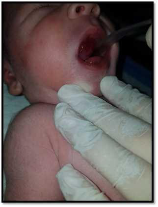
We herein present three cases of cystic hygroma that were admitted to our institution within a 4-month period. All of the three cases were identified as macrocystic, with two of them classified as giant cystic hygroma. All of the 3 cases presented to our neonatal intensive care unit within the first week of life for respiratory distress and required intubation for protection of airways. Beside the cystic mass, physical exam of all three newborns was unremarkable with no abnormal features. All of them were Syrian refugees with no appropriate prenatal care. Baby number 1 and 2 had a mass in the neck and underwent urgent aspiration to temporarily reduce the size of the cystic hygroma along with replacement therapy, awaiting surgical intervention. Unfortunately, both of them went into cardiac arrest few hours after the puncture and return of spontaneous circulation was not achieved. Baby number 3 had a mass in his mouth requiring recurrent aspiration for 3 weeks, however, he was a Syrian refugee and was lost to follow up with no information on his current medical status. Diagnosis for all of them was made clinically with characteristic cystic lesion that transilluminates. Ultrasound was made at the time of aspiration showing multicystic lesion with internal septations.
Cystic hygromas can manifest anywhere in the body, but most commonly occur as an isolated lesion on the neck, axillae or groin. They are usually encountered in the neonatal period or can present later, in early infancy. Patients can present with a painless, soft, asymptomatic visible mass that transilluminates or with complications.6 Congenital lymphangiomas are highly associated with chromosomal abnormalities, in up to 62% of cases, most commonly Turner syndrome.7 Thus, karyotype evaluation must be routinely done. Unfortunately, none of our patients had a karyotype done because of financial issues.
Although cystic hygromas are benign, complications can occur. Lesions can get infected, either by primary infection or secondary to nearby dissemination of microorganisms, leading to enlargement of the lesion, warmth and tenderness, which can progress to abscess formation. It can be associated with cellulitis and systemic illness. Treatment involves a course of antibiotics with drainage of abscess if present.6 Enlargement of the lesion can occur with systemic viral and bacterial infection. Spontaneous hemorrhage into the cyst is a frequently reported complication, requiring urgent surgical intervention. Other complications involve respiratory compromise and dysphagia secondary to compression from a neck mass.6 Severe respiratory distress may necessitate urgent tracheostomy. All of our patients had a mass in the head and neck region requiring intubation within the first week of life because of respiratory compromise (Figure 1, Figure 2).

Figure 1a and 1b Term babies (Baby 1 and 2), presented at birth with cystic hygroma of the neck with no antenatal diagnosis. Aspiration was done temporarily awaiting surgical excision; however, they passed away within few hours of aspiration because of cardiac arrest.

Figure 2 Baby 3, born term, presented for respiratory distress secondary to cystic hygroma of the mouth. He required recurrent aspiration of the hygroma.
Diagnosis can be made on physical examination at birth or later on during infancy or by prenatal ultrasonography. Ultrasound of the cystic mass shows multicystic lesion with internal septations and no blood flow on doppler. Other imaging techniques such as CT scan and MRI are important in case of surgical management to better delineate the lesion and its association with nearby nerves and vessels.8 MRI provides the most reliable diagnosis by demonstrating the full extent of complex lesions along with their macro- and microcystic components. They appear hyperintense on T2 sequences because of their high fluid content. Fluid-filled levels reflect layering of proteins and blood. Septae within the lesions are demonstrated by contrast enhancement.9 On histology, cystic hygromas are characterized by large, irregular vascular spaces lined with a single layer of endothelial cells with a fibroblastic, collagenous or fatty stroma, which may contain lymphoid aggregates, macrophages, smooth muscles or other local tissues. The cysts are filled with eosinophilic and proteinaceous fluid.10
Cystic hygromas are benign and can remain asymptomatic. However, treatment is required in case of complications and varies with the extent and location of lesions. Complete surgical excision remains the preferred treatment strategy, although it is a risky procedure. Extreme caution should be taken to avoid injury of nearby vessels and nerves. Complications include recurrence in 20% of completely excised lesions, wound infection, scar formation and incomplete resection.11 The recurrence rate is thought to be secondary to regrowth and reexpansion of incompletely excised lymphatic channels. Aspiration can be performed temporarily to reduce the size of the cystic hygroma, awaiting alternative intervention. A promising treatment modality that gained popularity in recent years is sclerotherapy with bleomycin or OK432, which has been documented to be successful in many case reports. Other treatment options with variable results include radiation, laser excision, radio-frequency ablation and cauterization. Within our experience, aspiration was associated with recurrence in all the cases. Non underwent surgical excision.
Cystic hygromas tend to be more extensive and harder to treat when they present earlier in life. The larger the lesion, the more it is associated with increased morbidity and mortality. Data in the literature regarding long-term follow up is still scarce and further prospective studies are needed to compare the effectiveness of different treatment modalities. Our 3 cases presented to our facility within a 4-month period 6 years ago and no other cases presented since then. All of the babies were Syrian refugees and two of them belonged to the same refugee camp. Can any environmental factors be related to this malformation? We leave the answer to future research.
None.
None.

©2022 Sacy, et al. This is an open access article distributed under the terms of the, which permits unrestricted use, distribution, and build upon your work non-commercially.