Journal of
eISSN: 2373-4426


A 12- year-old Caucasian girl presents with vaginal pain, redness and swelling of her vulva for 3 days. The onset was associated with fever, chills, and dysuria. She denies any sexual activity or trauma to the area. She has no history of sore throat, skin rash, arthralgia or arthritis, diarrhea, bloody stools, weight loss, abdominal or flank pain, frequency or urgency of urination. She denies ingestion of any prescribed or over the counter medications. The patient has no identified sick contacts. Her past medical history is unremarkable except for a previous episode of vaginal yeast infection 6 months ago.
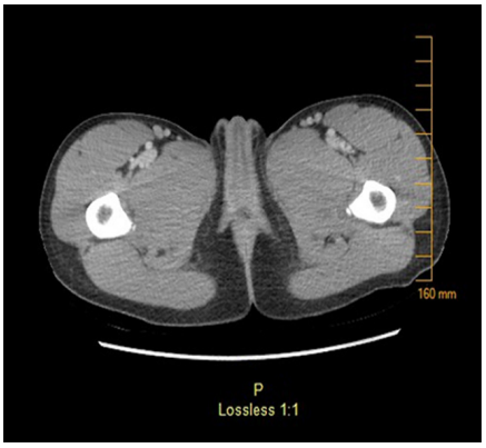
Physical examination reveals an anxious child, crying with pain. Her temperature is 38.7°C, heart rate is 109beats/min, blood pressure is 114/73 mm Hg, respiratory rate is 20 breaths/min, and oxygen saturation is 99% on room air. She has swelling and redness of clitoris, labia majora and labia minora with clear vaginal discharge observed on examination (Figure 1). There are bilateral ruptured vesicles on labia minora with dark discoloration of <1 cm on the right side. She also has moderate tenderness to palpation over the genital area, inner thighs, and bilateral enlarged inguinal lymph nodes. Her laboratory results are as follows: WBC count, 2.56×10³/mcL (2.56×10⁹/L) (52% neutrophils, 38% lymphocytes, 8% monocytes, 0.5% eosinophils); Hgb, 13.0 g/dL (133 g/L); Hct, 39% (0.39); and platelet count, 189×103/mcL (189×109/L). Her serum electrolytes and liver enzymes are normal. Her urinalysis obtained through a catheter shows clear yellow urine with negative leukocyte esterase and nitrite tests, 0 WBC/high power field (hpf), and 0 red blood cell/hpf. Viral PCR from genital lesions is negative for HSV 1 and 2. Serology for Cytomegalovirus (CMV) is negative for IgM and IgG. Epstein - Barr virus (EBV) mononuclear spot test and serology panel are negative for acute or past infection. ANA Screen Multiplex, ANCA Screen, and HCG Beta Subunit are all negative. A CT scan of the pelvis with contrast shows swelling and protrusion of the clitoral hood without evidence of any collection (Figure 2). A blood test reveals the diagnosis.

Figure 1 Physical examination shows swelling and redness of clitoris, labia majora and labia minora.

Figure 2 CT scan of the pelvis with contrast shows swelling and protrusion of the clitoral hood without evidence of any collection.
Clinical progression
The patient had Foley’s catheter placement due to pain and difficulty to urinate. She was started empirically on ampicillin-sulbactam and vancomycin for possible cellulitis, and awaiting bacterial cultures and laboratory test results. Vaginal swabs for bacterial cultures and nuclear acid amplification (NAA) for Chlamydia trachomatis, Neisseria gonorrhoeae, Trichmonas vaginalis, and Candida albicans were sent and were all negative. Both HSV 1&2 IgG antibodies in serum and Mycoplasma hominis PCR in urine tested negative. Antibiotics were discontinued after bacterial culture results reported no growth. The fever persisted for 1 week and the patient started to have transaminitis with ALT value of 219 U/L (normal, 0 to 55 U/L) and AST value of 268 U/L (normal, 5-34 U/L). The spleen was palpable 3 cm below the costal margin in the second week of illness. Quantitative EBV polymerase chain reaction (PCR) was sent in the blood and the patient had 12,800 copies/ml. EBV serology was repeated with a reported value of EBV VCA IgM antibody >4.0 (positive, >=1.1).
Differential diagnosis
Acute genital ulcers (AGU) in nonsexually active young adolescents have etiologies other than infections.1 Women with progesterone autoimmune dermatitis develop AGU that recur in the late luteal phase of their menstrual cycle when hormone levels are high. AGU were also reported in some pubertal premenarchal females or in the first year after menarche, which might be explained by high estrogen levels. Other etiologies like Crohn’s disease might present with knife cut vulvar ulcers or fissures that precede the gastrointestinal involvement. Additionally, Behcet’s disease manifests with AGU. It’s rare in USA but is common in Turkey and is seen worldwide. It is characterized by oral and genital ulcers that are painful and heal with scarring. Other manifestations of Behcet’s disease might include multi-organ inflammatory involvement in the form of uveitis, skin lesions such as erythema nodosum, and central nervous system involvement with the onset of genital ulcers. HIV, cyclic neutropenia, periodic fever, aphthous stomatitis, pharyngitis, adenitis (PFAPA) syndrome, and mouth and genital ulcers with inflamed cartilage (MAGIC) syndrome are part of the differential diagnosis of AGU but patients tend to develop recurrent ulcers with other systemic manifestations.
Final diagnosis
The diagnosis was Lipschutz ulcers secondary to primary EBV infection.2
In 1913, Lipschutz reported a series of young virgin girls who developed vulvar ulcers without a definite infectious source. The term “Lipschutz ulcer” is used to refer to vulvar or lower vaginal ulcers with no identifiable etiology, based on clinical, histopathologic and direct cytotoxic microbiologic findings. It is also known as “ulcus vulvae acutum” or “acute genital ulcer”.
AGU in nonsexually active young adolescent girls occur rarely. The precise incidence is not known but the average age at onset was 12 to 29 years based on a few published case series. The etiology of non-venereal AGU have been attributed to different causes including autoimmune, infectious, and idiopathic etiologies. Over 70% of all reported patients with non-venereal AGU were diagnosed with idiopathic etiology while primary EBV infection has been cited as the commonest infectious cause found in nonsexually active girls. In 25 reported cases with AGU, primary EBV infection was diagnosed by serologic testing mostly, or from the genital ulcers using viral culture or polymerase chain reaction. This suggests that EBV infection can cause direct cytotoxicity or elicit an immunologic response that manifests as vulvar ulcers. Other infectious etiologies included CMV, mycoplasma, influenza A&B, mumps, paratyphoid fever and salmonella.
Lipschutz ulcers present as single or multiple ulcers with raised demarcated borders. Many lesions will develop gray pseudomembrane or adherent brown eschar with secondary erythema, edema, and inguinal lymphadenopathy. Our patient had significant clitoral enlargement which was not reported previously in the literature. Usually, ulcers are distributed on the medial aspects of the labia minora, but they might be found on the labia majora, perineum, and in the lower vagina. “Kissing” ulcers on opposing labial surfaces are not uncommon. Size of ulcers is variable but lesions can measure greater than 1 cm in diameter. Most of ulcers resolve with complete healing. Around 71% of reported patients in the literature presented with systemic symptoms in the form of fever, myalgia, and fatigability. In addition, 47% of cases had a history of oral ulcers, and around 30% were reported to have recurrent episodes of AGU.
The laboratory evaluation of AGU should include a viral HSV PCR 1&2 of the lesion to exclude sexually transmitted infection. Viral culture for HSV has a limited sensitivity. Diagnostic tests for syphilis, chancroid, and lymphogranuloma venereum might be considered in the initial work up depending on the prevalence and epidemiology of these infections. A complete blood count will help in detecting, anemia, thrombocytopenia, and neutropenia. Serologic testing for EBV is highly indicated. Negative serology for EBV with AGU does not exclude a primary infection with EBV since genital ulcers might proceed the onset of other systemic manifestations. Detection of EBV DNA by PCR in serum or directly from the ulcer or vulval swabs is diagnostic. Serology for CMV IgM and IgG is recommended. Iron, folate, Vitamin B12 levels, and a biopsy of the vulvar lesion are indicated for patients with recurrent AGU without identifiable etiology.
Treatment of non-venereal AGU is mainly supportive. Sitz baths and a mixture of lidocaine, epinephrine and tetracaine might be helpful in controlling local pain in AGU. Alternating topical corticosteroid ointment or amlexanox paste with a local anesthetic are reported to be helpful. Antibiotics are not indicated except if secondary cellulitis develops during the acute onset of genital ulcers which is reported in 30% of the cases.
Most of non-venereal AGU heal within 16-21 days. EBV or CMV serology can be repeated especially if the initial test was negative. Evidence of seroconversion supports the diagnosis of an acute primary infection and carries a lower risk of recurrence of AGU. It is important for patients to follow up yearly with their primary care providers for ruling out progression to other systemic manifestations that will change the diagnosis.
The patient’s vulvar swelling improved significantly throughout the admission, and Foley’s catheter was removed 2 days prior to discharge. The pain medications were tapered off to oral acetaminophen. She was instructed to use topical corticosteroid ointment and zinc oxide for barrier cream. She was discharged from the hospital on day 14. The patient’s ulcers healed completely and the liver enzymes dropped to normal levels within four weeks of the onset of illness.
Lessons for the clinician
None.
I have no financial disclosures related to this article to disclose.
I have no conflicts of interest to disclose.
None.

© . This is an open access article distributed under the terms of the, which permits unrestricted use, distribution, and build upon your work non-commercially.