Journal of
eISSN: 2379-6359


Review Article Volume 13 Issue 3
1Senior Resident ENT, GSMCH, India
2Professor & Head ENT, GSMCH, India
3Professor Oral & Maxillo-Facial Surgeon, Dy Patil University, India
Correspondence: Dr. Mohit Srivastava, Professor & Head ENT, GSMCH , Hapur, House No 1153, 2nd Floor , Sector -5 Mohan Mekins Society Vasundhara, Ghaziabad, India, 201012, Tel +91 9999556665
Received: September 15, 2021 | Published: October 5, 2021
Citation: Gupta K, Srivastava M, Singh V. Rhino-orbito-cerebral mucormycosis (black fungus) in covid 19 patients in western Uttar pradesh India. J Otolaryngol ENT Res. 2021;13(3):79-82. DOI: 10.15406/joentr.2021.13.00495
Mucormycosis (Black fungus) is a designated as a rare, rapidly progressive fatal disease of immunocompromised caused by saprophytic fungus of family mucorales. Early diagnosis with prompt medical and surgical treatment is the only tool available. Rhino-orbito-cerebral is the most common subtype. In India we saw a sudden rise in mucormycosis cases during second wave of COVID 19. This necessitated a systematic review of epidemic of mucormycosis in COVID 19. A Retrospective multi-centric study was conducted comprising of 51 cases of Rhino-orbito-cerebral mucormycosis with present or recent COVID19 in Western Uttar Pradesh positive status presenting to us during 14th April 2021- 31st May 2021. Either Type2 Diabetes Mellitus or history of recent use of steroids in high doses was present in all the patients. Contribution of virulence of the Delta strain B1.617.2 is significant. FESS with sino-nasal debridement contributes significantly towards mortality reduction and cost of total treatment by significantly reducing days of Liposomal Amphotericin B therapy. Early diagnosis with prompt medical and surgical management along with blood sugar control and avoiding use of high dose of steroids remain to key to mortality and morbidity reduction.
Keywords: black fungus, mucor, mucormycosis, rhino-orbito-cerebral, causes, treatment, covid 19, india, sugar, steroids, steam, oxygen, surgery
Mucormycosis are a group of invasive infections caused by filamentous fungi of the Mucoraceae family.1 It is the third invasive mycosis in order of importance after candidiasis and aspergillosis.2 The incidence of mucormycosis is approximately 1.7 cases per 1000000 inhabitants per year, and the main risk-factors for the development of mucormycosis are ketoacidosis (diabetic or other), iatrogenic immunosuppression, use of corticosteroids or deferoxamine, disruption of mucocutaneius barriers by catheters and other devices, and exposure to bandages contaminated by these fungi.2
Rhino-orbito-cerebral is the most common clinical subtype of disease. Mucormycosis is a difficult to diagnose rare disease with high morbidity and mortality.3 This form presents with sinusitis, facial and eye pain, proptosis, progressing to signs of orbital structure involvement.4-7 Necrotic tissue can be seen on nasal turbinates, septum and palate. This may look like a black eschar.7,8 Intracranial involvement develops as the fungus progresses through either the ophthalmic artery, the superior fissure, or the cribiform plate.4,7
Diagnosis of mucormycosis rests upon the presence of predisposing conditions, signs and symptoms of disease, observation of fungal elements of specific morphology in histological sections, and direct smears of material, and, to a lesser extent, culture results.6,7 There are no reliable serological tests for diagnosis at present.8 The incidence of mucormycosis has risen more rapidly during the second wave compared with the first wave of COVID 19 in Western Uttar Pradesh India, with atleast 28,252 mucormycosis cases on 7th June 2021. 86% of them are known to have history of COVID 19 and 62.3% of them are known to be diabetic.9
To study various risk factors, clinical features, diagnosis, treatment and outcome of mucormycosis patients during second wave of COVID 19 in Western Uttar Pradesh India.
Study design
Multi-centric Retrospective.
Sample size
No of cases- 51
Study period- 14th April 2021- 31st May 2021.
Inclusion criteria
All the following criteria was satisfied
Patient presented during 14th April 2021 midnight- 31st May 2021 midnight.
COVID 19 RT PCR positive at any time during the study period or within 28 days before beginning of study period.
Biopsy proven mucormycosis and/or patient had features clinically consistent with diagnosis of mucormycosis, that is, two or more of following on presentation:
Black eschar within oral cavity and/or blackish eschar within nasal cavity and/or blackish eschar over face.
Severe facial pain and facial swelling of onset within last 28 days.
Eye swelling and/or ptosis and/or proptosis.
Computerised tomography or magnetic resonance and imaging suggestive of invasive fungal rhinosinusitis.
Exclusion criteria
Oral and sino-nasal malignancies, other conditions associated with oro-mucosal ulcerations, absence of present or recent COVID 19 status.
After all inclusion and exclusion criteria were satisfied, records were checked for presence and absence of various predisposing factors, treatment offered, histopathology reports, surgeries performed and outcome. All the data was gathered and tabulated in Microsoft Excel 2008 spreadsheet. SPSS 24 was used for statistical calculations. Results were systemized and summarized.
Observations
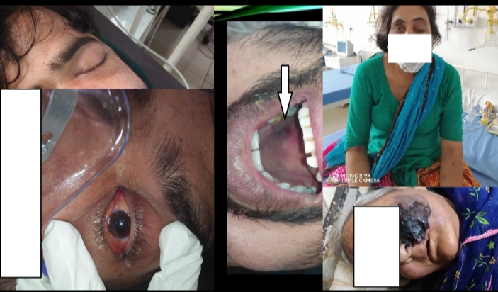
(Tables 1.1-1.6), (Images 1.1-1.3).
|
Sex |
No of cases |
|
Male |
28 |
|
Female |
23 |
|
Total |
51 |
Table 1.1 Sex distribution of cases
|
Age distribution |
No of cases |
|
Less than 31 |
2 |
|
31-45 |
22 |
|
46-60 |
18 |
|
More than 60 |
9 |
|
Total |
51 |
Table 1.2 Age distribution of cases
|
COVID Status |
No of cases |
|
Active COVID |
25 |
|
Post COVID |
26 |
|
Total |
51 |
Table 1.3 COVID status of patients
|
Risk factor |
No of cases |
Association |
|
Diabetic |
43 |
Strong |
|
Recent history of Steroids |
49 |
Strong |
|
Either Diabetes or steroids |
51 |
Definitive |
|
Oxygen support |
16 |
Weak |
|
History of Tocilizumab |
zero |
Can not comment |
|
Steam inhalation more than one hour a day |
1 |
Absent |
Table 1.4 Various Risk Factors for mucormycosis with Delta stain of COVID 19 noted in our study
|
Clinical feature |
No of cases |
Frequency |
|
Eye swelling |
34 |
66.67 |
|
Diminished vision |
29 |
56.86 |
|
Ptosis |
28 |
54.9 |
|
Black eschar |
25 |
49.02 |
|
Facial swelling |
23 |
45.1 |
|
Proptosis |
18 |
35.29 |
|
Facial pain |
16 |
31.37 |
|
Loss of vision |
11 |
21.57 |
|
Nasal discharge |
8 |
15.69 |
|
Nasal bleed |
4 |
7.84 |
|
Altered sensorium |
1 |
1.96 |
Table 1.5 Clinical features in 51 patients of Mucormycosis with COVID 19
|
Outcome |
No of patients |
Frequency |
|
Recovered during study period |
18 |
35.29 |
|
Survived but did not recover during study period |
23 |
45.1 |
|
Facial disfigurement |
4 |
7.84 |
|
Permanent loss of vision from one eye |
3 |
5.88 |
|
Permanent loss of vision from both eyes |
1 |
1.96 |
|
Expired during study period |
10 |
19.61 |
Table 1.6 Outcome in 51 patients of Mucormycosis with COVID19
|
Treatment given |
No of patients |
Frequency |
|
Liposomal Amphotericin B for 1-7 days |
35 |
68.63 |
|
Liposomal Amphotericin B for 8-14 days |
11 |
21.57 |
|
Liposomal Amphotericin B for more than 14 days |
1 |
1.96 |
|
Debridement surgery |
34 |
66.67 |
Table 1.7 Treatment offered in terms of Liposomal Amphotericin B and Debridement surgery to various patients
|
Surgery and survival |
No of patients who took surgery |
No of patients who did not take durgery |
Total patients |
|
No of patients survived during study period |
32 |
9 |
41 |
|
No of patients expired during study period |
2 |
8 |
10 |
|
Total patients |
34 |
17 |
51 |
Table 1.8 Number of patients who took surgery charted with number of patients who survived the study period
|
Duration of amphotericin B therapy |
1-45 days |
|
With Surgery |
1-7 days |
|
Without Surgery |
1-45 days |
Table 1.9 Effect of surgery on duration of Amphotericin B therapy and hence cost of treatment
|
Duration from presentation |
No of patients who expired |
|
Within 24 hours |
4 |
|
24-48 hours |
2 |
|
48-72 hours |
1 |
|
More than 120 hours |
3 |
|
Total |
10 |
Table 1.10 Figures of mortality from the time of presentation

Image 1.1 Clinical features of mucormycosis. Clockwise: Facial swelling, oral cavity eschar, eye swelling, facial eschar, congestion, diminished vision and ptosis of eyes.
The disease is equally seen in both sexes. The disease is exclusively seen in either diabetics or those who have recently taken steroids. Immuno-compromised patients with Delta stain of COVID 19 Pango lineage B.1.617.2 have more risk of developing mucormycosis than their non COVID counterparts. Oxygen inhalation also contributes to the risk. There is no positive or negative effect of steam inhalation.
There is an increase in no of cases of mucormycosis because of delta strain of COVID 19. There is a shift of peak towards the younger age groups. There is increased frequency of eye involvement. Mortality is maximum within first 72 hours of presentation. However, mortality ratio is less when co-infected with delta strain of COVID 19.
Surgery offers significant benefit by decreasing mortality, decreasing duration of Liposomal Amphotericin B treatment and hence reducing cost of treatment.
Delta strain of COVID 19 Western Uttar Pradesh India has significantly increased the incidence of mucormycosis due to its immunosuppressive effect. Excessive use of steroids has also contributed to the same. Since, patients of younger age group are affected more with this strain, the peak of mucormycosis has also shifted in the same direction. In a young patient with unatherosclerosed and more patent vessels, there is early involvement of ethmoid and ophthalmic vessels and hence early necrosis of turbinate’s is seen along with prominent eye symptoms. The massive coverage of black fungus by media has made people extra conscious about the mucormycosis, which has also contributed to early presentation and early diagnosis of the disease. Early diagnosis coupled with early surgery in younger patient may have contributed to lower mortality when recorded over a short time span.
None.
None.
None.

©2021 Gupta, et al. This is an open access article distributed under the terms of the, which permits unrestricted use, distribution, and build upon your work non-commercially.