Journal of
eISSN: 2373-6410


Case Report Volume 12 Issue 4
1Independent clinical neurology, Colombia
2Epidemiologist, Universidad el Bosque, Colombia
3Pediatric Computational Imaging Center, Hospital Sant Joan de Déu, Spain and Centro de Investigación Biomédica en Red, Spain
Correspondence: Andrea Catalina Nassar, Independent clinical neurology, Carrera 21A # 124-55, Colombia
Received: July 26, 2022 | Published: August 11, 2022
Citation: Nassar AC, Peña FY, Stephan-Otto C. Brain volumetric changes after sensory isolation: single case experimental design. J Neurol Stroke. 2022;12(4):114-117. DOI: 10.15406/jnsk.2022.12.00512
This study aimed to investigate the cerebral volumetric changes measurable by brain magnetic resonance imaging, as well as the different physiological variables documented through neurobiofeedback equipment, before and after 30 serial sessions of guided meditations in a sensory isolation tank. A pilot test was carried out, with a single subject, and it was concluded that serial sessions of meditations in flotation tanks changed the brain structure, triggered the relaxation response, generating greater physical, psychological and mental well-being for the research participant.
Keywords: sensory isolation tank, mind body medicine, relaxation response, stress, neurobiofeedback, longitudinal magnetic resonance case study
In recent years, it has been more frequently documented that many pathophysiological conditions are related to emotional disorders such as depression, anxiety and especially stress. The last has been defined as a feeling of physical tension or emotional that can originate in a specific situation or in thoughts of frustration, anger, and irritability.1,2
It is also considered that the stress response is an innate situation that is associated with the need for survival known as the fight-or-flight response; Regarding the positive response to stress determined in a survival reaction, while the negative response is one that is maintained under conditions where there is no precise justification for survival and on the contrary the relaxation response (RR) has been dismissed, which provides a reaction of tranquility, peace, and inner harmony.3 On this last case, when the stress response persistently predominates, it is called allostatic overload;4 In addition, there may be an increase in limbic activity, as well as cortisol levels, which in turn triggers systemic neuroimmunoendocrine disorders harmful to health and there is even evidence of a shortening of chromosomal telomeres inducing premature aging among other changes harmful to health.5
In order to counteract the negative effects of stress and associated adverse situations such as depression and anxiety, it has been observed that the induction of a RR in a structured and replicable way, can considerably reduce the pathophysiological effect of stress on the body.6,7 On the other hand, one of the most surprising findings that has been reported is that the RR, as well as other techniques that induce peace and inner tranquility, indicate that it can achieve structural anatomical changes in the human brain, measurable by magnetic resonance (MR).8,9 To induce an appropriate RR, it has been recommended to have sensory deprivation, in order to create a harmonious environment where the individual can self-induce positive thoughts and of tranquility; a known effective way to induce sensory deprivation are the flotation tanks (FT), as indicated Kjellgren y Westman (2014) cited by Rey (2015).10
During the Float-REST method (Restricted Environmental Stimulation Technique) the individual is disposed in a horizontal posture inside the flotation tank with water containing highly concentrated salt of magnesium sulfate. All incoming stimuli are reduced to a minimum for a period of approximately 45 minutes. During this time there are no sounds, no light, and the water in the tank is at a normal body temperature of approximately 37 degrees Celsius. This method has been scientifically researched and is considered today a proven and effective way to reduce stress, depression, anxiety, and increase optimism and the quality of sleep, and in some cases even significant pain reduction has been reported.10
FTs have been used as catalysts and potentiators in the treatment of different pathologies, in combination with relaxation and meditation techniques; These last two are also used in mind-body medicine3 as inducers of the RR and have been associated with important cerebral volumetric changes measured by means of MRI of structures such as the amygdala, insula, hippocampus, and the prefrontal cortex, among others. These areas are associated with the regulation of emotions, memory, attention, decision making, learning, empathy, compassion, and self-awareness, which can influence the physical, mental, emotional, and spiritual well-being of people.11,12 Given the importance of these techniques, it was considered pertinent to investigate whether there are brain anatomical changes measurable by MRI after inducing an appropriate RR by effective sensory deprivation in TF.
A longitudinal descriptive pilot study was carried out between October 2018 and March 2019, whereby an experimental test a single participant without neuropsychiatric pathology was selected, of legal age, who had not been exposed to sensory isolation in TF during the last three months. The participant signed the informed consent and voluntarily accepted the conditions established, complying with the requirements and ethical standards of research in human beings.
Study variables and instruments: Sociodemographic information was obtained, such as age, sex, marital status, number of children, mind-body techniques practiced, and later the activities to be carried out were explained in detail. A neurological and psychiatric evaluation by specialists was also performed. The participant was then taken to a TF immersion training workshop, and another workshop on guided meditation; A nuclear magnetic resonance image of the brain was taken with the Magnetom essenza D12 serial 3.T 72021 (Germany) equipment. The participant was then exposed to 30 sessions of sensory isolation under guided meditation in a Samadhi flotation tank (USA). Before and after each TF immersion, a neurobiofeedback session was performed in order to assess whether a relaxation response was produced, for a total of 60 sessions with the Biofeedback equipment H003 Multi-channel GP8, with 8 low-noise universal channels (Washington, USA). At the end of 30 days, another brain MR image was taken in order to show structural anatomical changes. All the information was taken to a database for later analysis.
Data analysis: The data obtained through neurobiofeedback in relation to brain waves, heart rate, respiratory rate, muscle tone, temperature, and galvanic skin response, before and after guided meditation on the TF, were analyzed using the specific software SPSS version 20.0. Structural data extracted from brain MR images were automatically processed using FreeSurfer version 6.0, for cortical reconstruction and segmentation. Standard processing was done to extract reliable brain volume estimates, at the level of the following brain structures: right amygdala, left postcentral gyrus, left rectus gyrus, right medial frontal region, left subcallosal area, left superior parietal lobe, left entorhinal area, right globus pallidus, and left amygdala. Brain volumes were compared before and at the end of the study.
Ethical considerations: The study was approved by the ethics committee of the Central Military Hospital in Bogotá, Colombia. The investigators declared to be familiar with the norms for research in human beings based on the Nuremberg Code, the Belmont Report, and the Declaration of Helsinki. In accordance with Resolution 8430 for research on human beings in Colombia, this was an investigation, with risk greater than the minimum, due to the participant underwent two brain magnetic resonance studies, required multiple TF immersions. and the performance of 60 neurobiofeedback sessions. The participant agreed to participate and signed the informed consent.
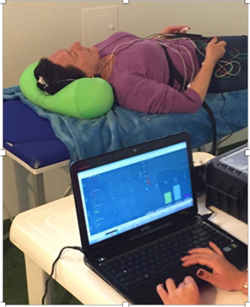
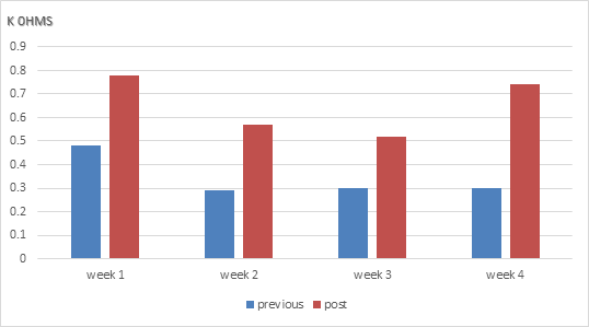
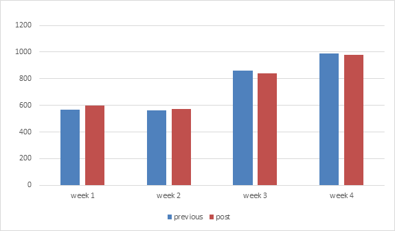
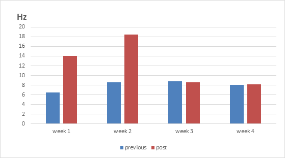
The pilot test was carried out with a single female participant, 63 years old, with no history of comorbidities (Figure 1). In relation to the physiological variables, registered by means of the neurobiofeedback equipment throughout the 30 days of the investigation, a decrease in average heart rate was observed after immersion in the TF (Figure 2) and an increase in both body temperature and galvanic resistance and Alpha wave activity. (Figures 3–5). The weekly average number of breaths was very similar, being slightly higher in the first two weeks and slightly lower in the last two weeks after immersion in the TF (Figure 6); On the other hand, the average electromyographic activity increased in the first two weeks after the immersion. and remained very similar to the average before the emersion in the last two weeks (Figure 7).

Figure 1 Photograph showing the moment of the neurobiofeeback test performed on the participant before and after immersion in the flotation tank.

Figure 4 Average of the galvanic resistance of the skin before and after immersion in a flotation tank.

Figure 6 Average number of respirations during the neurobiofeedback test before and after immersion in flotation tanks.

Figure 7 Average electromyographic change before and after immersion in a flotation tank. Regarding cerebral volumetric changes.
Regarding cerebral volumetric changes, an increase pattern was observed in the gray matter, as well as an increase in the volume of the right amygdala, left postcentral Gyrus, left postcentral gyrus, left rectus gyrus, right medial frontal region, left subcallosal area, left superior parietal lobe, left entorhinal area, right globus pallidus, left amygdala. The volumetric changes ranged from 3% to 10% (Figure 8), expected findings for the proposed hypothesis.
In this study it was possible to evaluate by the cerebral magnetic resonance, changes in the volume of different brain structures, as well as determining changes at the level of brain waves, heart rate, respiratory rate, muscle tone, temperature, and the galvanic response of the skin. It was found that after 30 consecutive days of practicing a meditation within a TF, brain changes occurred at the level of structures such as the right amygdala, left postcentral gyrus, left rectus gyrus, right medial frontal region, left subcallosal area, left superior parietal lobe, left entorhinal area, right globus pallidus, and left amygdala. Likewise, the relaxation response was triggered by evidence through neurobiofeedback changes in blood pressure, heart rate, muscle tone, galvanic skin response, temperature, and brain alpha waves, which indicates that a well-being was generated at the physical, psychological, and mental level of the participant. On the other hand, it should be considered that the evidence supporting the importance of the experiential influence on the development of the nervous system is increasing, especially sensory deprivation studies, which have contributed to the understanding of the pathophysiological phenomena underlying alterations in a superior cognitive function.13
For several decades, TFs have been used to decrease visual, auditory, olfactory, thermal, tactile, gravitational, proprioceptive cues, as well as movement and language to trigger adequate RR.14 Some of the work in this context by Turner and Fine of the Medical College of Ohio documented that floating in TF activated the RR and counteracted the effects of the fight-or-flight response.14
Different groups of researchers have carried out studies, where they use the tank as part of the treatment of different pathologies. Most of the studies have been carried out on different anxiety disorders, panic attacks, chronic pain, headache, sleep disturbances and major depression, among others. In the different investigations it was evidenced that the tank is reliable, safe, and that it decreases the frequency, duration and intensity of the symptoms of the different pathologies.15–22 Thus, studies such as those by Bood;15 Bood, Sundequist, Kjellgren, Nordström and Norlandejem (2005) and Kjellgren22 cited by Kjellgren and collaborators,17 indicate that treatments in a flotation ion tank (Flotation-Rest) with relaxation, can generate a multitude of positive effects such as stress reduction, significant reduction in stress-related pain, the increase in optimism and decrease in the degree of depression and anxiety, which are mainly mediated by deep relaxation.17
This relaxation response is evaluated using neurobiofeedback methods where an electrical device measures intracorporeal variation, allowing precise evaluation of different physiological variables such as brain waves, muscle tone, galvanic response of the skin, temperature, heart rate and respiratory rate, to verify whether this state of relaxation has been triggered.3
Several studies have shown by means of magnetic resonance brain structural and electroencephalographic changes that occur during and after meditations;5,6 These researchers showed that different meditation techniques can change the size and function of some brain regions such as the amygdala, the insula, the hippocampus and the prefrontal cortex, among others; areas that are associated with emotion regulation, memory, attention, decision-making, learning, empathy, compassion, and self-awareness.11,12,23,24 Additionally, studies that have used neuroimaging and electroencephalography to assess the proposal of meditation, show structural and functional changes in sectors such as the prefrontal cortex, insula, hippocampus, cingulum, primary visual cortex, and the striatum.25
These results are reaffirmed by what was exposed in an investigation on the influence of mental silence on the human brain led by the Universidad de La Laguna, which determined that people who meditate have a greater volume of gray matter throughout the brain with the greatest significant differences in the right hemisphere: right temporal, right frontal, and right brainstem. Furthermore, the six areas with the greatest significant differences were also in the right hemisphere: middle and inferior temporal lobe, orbital and inferior frontal cortices, paracentral and precentral lobes.26 Other authors have described the importance of the practice of meditation in the healing of traumatic emotional aspects associated with conflicts intrinsic to personality, as well as an essential part for an integral health of the individual.27
Additional studies found a higher density of gray matter in the hippocampus, an important area for learning and memory, as well as in structures associated with self-awareness, compassion, and introspection. In addition to the stress reduction reported by the participant in this study, it correlated with lower gray matter density in the amygdala, an area that plays an important role in anxiety and stress. It is considered pertinent to extend these results to a larger series of participants to show the response in different individuals and possibly better categorize its usefulness according to other physical, emotional, mental, and intellectual variables.28,29
These findings open the doors to innumerable possibilities for future research on the potential of the relaxation response as a treatment for stress and other disorders related to psychological and psychosomatic pathologies.
Serial meditation sessions in flotation tanks can change brain structure, trigger a relaxation response, and lead to greater physical, psychological, and mental well-being.
Special Thanks Centro de Flotación Gravedad Cero, Bogotá, Colombia.
The authors declare that there is no conflict of interest for the preparation or publication of this documents.

©2022 Nassar, et al. This is an open access article distributed under the terms of the, which permits unrestricted use, distribution, and build upon your work non-commercially.