Journal of
eISSN: 2373-6410


Case Report Volume 6 Issue 6
1Department of Neurosurgery and Spinal Surgery, Brandenburg Medical School, Campus Neuruppin, Germany
2Department of Neurosurgery, University Hospital of Udine, Italy
3Department of Neurosurgery, Kantonsspital Winterthur, Switzerland
Correspondence: Alex Alfieri, Chairman, Neurological and Spinal Surgery, Brandenburg Medical School Campus Neuruppin, Fehrbelliner Straße 38 D-16816 Neuruppin, Germany, Tel +49 (0)3391 39-47600, Fax +49 (0)3391 39-47609
Received: February 02, 2017 | Published: May 22, 2017
Citation: Yaish M, Török L, Zanotti B, Alfieri A (2017) Familial Colloid Cyst of the Third Ventricle in Monozygotic Twins. J Neurol Stroke 6(6): 00219. DOI: 10.15406/jnsk.2017.06.00219
Colloid cysts are slow-growing non-neoplastic true epithelium lined cysts of the central neuraxis, usually arising in the third ventricle.1 The first description was published in 1858 by Wallmann2 as an autopsy finding. They constitute 0.5-2% of all intracranial tumors and comprise 55% of the third ventricle’s lesions.3 Colloid cysts are and can be found in patients of any age, with a peak between the second and the fourth decades, and no differences in distribution according to sex.4 The wall usually consists of a collagenous connective tissue stroma lined by single layer of columnar epithelium.4
Colloid cysts are often found incidentally, but when symptomatic, they present usually with signs and symptoms of acute or chronic obstructive hydrocephalus, by occlusion of the foramen of Monro.5 Sudden death, as consequence of untreated hydrocephalus and raised intracranial pressure, is well recognized in patients with colloid cyst.4˗6 The treatment is surgical, endoscopic or microsurgical.4
A study of 155 patients with newly diagnosed colloid cysts showed four factors associated with cyst related symptoms: 1) younger patient age (44 years versus 57 years), 2) cyst size (13 mm versus 8 mm), 3) ventricular dilation (83% versus 31%),and 4) increased signal on T2-weighted magnetic resonance images (44% versus 8%).1 More than half of cases were discovered incidentally. In five patients with incidental cysts (8.8%) an enlargement of the cyst was documented and a surgical procedure was done. Nearly half of symptomatic patients present hydrocephalus, more than 10% present an acute symptom, and 1% of patients decease due to obstructive hydrocephalus and herniation.1
Some familial cases were previously reported, including monozygotic and dizygotic twins. Most of the familial colloid cysts were treated operatively, although few cases were treated conservatively, with regular radiological follow up.7˗9 To our knowledge, only 4 pairs of twins with colloid cyst have been previously reported in the literature, three of which are monozygotic and the other one is dizygotic.10˗13
In this report, we present the fourth monozygotic twins with this potentially life-threatening pathology. The two brothers were operated four years apart after presenting with different clinical scenarios.
Brother A
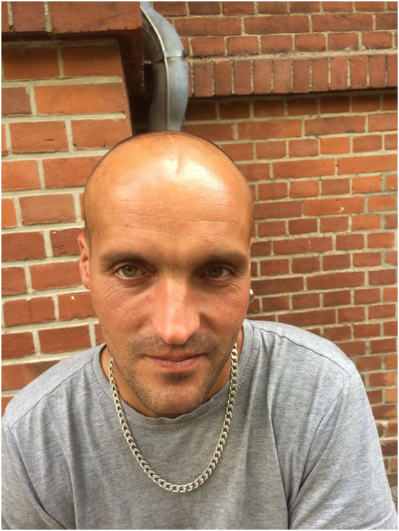
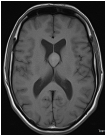
The 31-years-old (Figure 1) patient was admitted to a hospital in another country in 2012 following a generalized tonic-clonic seizure. After first stabilization the patient was neurologically intact without any signs and symptoms of increased intracranial pressure. A prophylactic antiepileptic therapy with levitracetam was initiated. An MRI was done and showed an isointense in T1 and T2WI and a hyperintense mass in the fluid attenuated inversion recovery sequence suggesting the diagnosis of a colloidal cyst of the third ventricle causing the pathognomonic obstructive hydrocephalus involving only the lateral ventricles (Figures 2-5).

Figure 1 Brother A, year 2016. The microsurgical removal of the colloid cyst was performed in year 2013. Permission kindly granted by the patient.

Figure 2 MRI T1 (Brother A): 18x22 mm well circumscribed colloid cyst in the 3rd ventricle extending in the lateral ventricles.
The cyst was removed through a right frontal craniotomy through a transcortical transventricular approach. Postoperatively, the patient was discharged after two weeks with an intact neurological status. Because the patient was in another country, no further investigations were done.
Brother B
In 2016, while working as a carpenter, monozygotic brother B (Figure 6) suddenly fell into a generalized tonic-clonic seizure. When the emergency doctor arrived at the workplace the patient presented a Glasgow Coma Score of 4 (E1, V1, M2), wide bilateral pupils without response to the light. The patients was intubated and conducted to the emergency room of our hospital. The neurologic status offered no brainstem reactions, wide pupils bilateral without response to the light, no motor response. Funduscopic examination revealed acute papilledema. A non-contrast CT of the brain showed a well-defined 30x33 mm hyperdense lesion in the third ventricle with massive obstructive hydrocephalus of the lateral ventricles (Figure 7), with the classical appearance of a colloid cyst. In further questioning, the family reported that the patient suffered a history of headache with nausea and vomiting over the last week. Due to the wrong neurological status of the patient an MRI was omitted preoperatively and the patient was urgently microsurgically treated. Under general anesthesia, a frontal craniotomy was made in the right side in supine position and through a transcortical-transventricular approach the cyst was microsurgical removed with a temporary placement of a closed external ventricular drain (EVD) to prevent the possible complication of hydrocephalus. Postoperatively, the patient was early extubated and he was neurologically intact. The pupils were bilateral isochoric, miotic and well responsible to the light. During the first three days postoperatively there was not need to open the EVD, and consequentially it was removed. The histopathological analysis confirmed the diagnosis of colloid cyst, which was positive for CK AE1/AE3 and EMA, negative for CDX-2, TTF-1 and S-100. A 5 days follow up CT showed a mild hydrocephalus (Figure 8). The patient was discharged in the second postoperative week without neurological deficit. A neuropsychological evaluation after six months revealed mild attention impairments. The patient went back to work after six months. The rest of the family underwent prophylactic screening without other pathological signs.
Familial colloid cysts are extremely rare; only around 30 cases were mentioned in the literature.7˗13 With the presented cases there will be until now 5 registered pairs of twins with colloid cysts with a total of four monozygotic twins and one dizygotic twin. The autosomal dominant inheritance was previously postulated.9 These findings suggest an increase in the incidence of colloid cyst in twins as compared to the general population. Accordingly the performing of a magnetic resonance imaging in both twins is advisable, if one of them was proved to have a colloid cyst.
Treatment options include conservative, microsurgical, or endoscopic treatment.4,14,15 An asymptomatic patient may be managed with follow-up imaging studies.1 If neurological signs are seen, prompt imaging studies should be obtained and a timely microsurgical or endoscopic resection planned.4,14,15 In case of brother B, the rapid clinical deterioration has forced an emergency operation without performing further diagnosis. A further knowledge from this case is that, although brother B was admitted with wide pupils and without brainstem reflexes, the timely operation allowed a prompt weaning, and the patients presented no neurological losses. The slightly attentional impairments should be observed in the long time follow-up. According to these findings, a genetic origin of colloid cysts can be supposed. Further genetic studies are needed to confirm these observations.
None.
None.
None.

©2017 Yaish, et al. This is an open access article distributed under the terms of the, which permits unrestricted use, distribution, and build upon your work non-commercially.