Journal of
eISSN: 2377-4282


Research Article Volume 5 Issue 6
1Department of Medical Physics, Bharathidasan University, India
2Department of Chemistry, Sree Sevugan Annamalai College, India
Correspondence: Nehru LC, Department of Medical Physics, School of Physics, Bharathidasan University, Tiruchirappalli-24, Tamilnadu, India
Received: June 14, 2017 | Published: July 21, 2017
Citation: Hariharan D, Srinivasan K, Nehru LC (2017) Synthesis and Characterization of Tio 2 Nanoparticles Using Cynodon Dactylon Leaf Extract for Antibacterial and Anticancer (A549 Cell Lines) Activity. J Nanomed Res 5(6): 00138. DOI: 10.15406/jnmr.2017.05.00138
The synthetic techniques, for the synthesis of Metal oxide nanoparticles involve toxic solvents, high pressure, high-energy and high temperature which may be efficient but not eco-friendly. The synthetic techniques for metal oxide nanoparticles using plants, has several advantages over the chemical synthesis such as cost effectiveness, simplicity as well as compatibility for medical applications. In this part of the study we are using the cynodon dactylon leaf extracts to synthesize titanium dioxide nanoparticles. The properties of TiO2 include high refractive index, light absorption, non toxicity, chemical stability and relatively low cost production. The cynodon dactylon leaf has reduced the size of TiO2 nanoparticles. In the present study antibacterial effect of TiO2 nanoparticles on E. coli strain was analyzed. The well diffusion assay is used to confirm the inhibition zone of bacteria. Further the characteristics of the obtained TiO2 nanoparticles were studied using XRD, FTIR, Laser Raman spectroscopy, Scanning Electron Microscopic and the results are presented in detail.
Keywords: tio2, cynodon dactylon, antimicrobial activity
The nano particles are different from the large particles of the same composition because of their large surface area and volume ratio. The metal oxide nanoparticles have received considerable attention on medical line due to their antibacterial properties, resistance against microbes, drug delivery, antibiotics and immune chromatography, tissue / tumour image, anticancer activities and identification of pathogens in clinical specimens.1˗2 TiO2 is the most promising material in the group of the metal oxides. In n - type semiconductor, titanium dioxide is a most important semiconductor due to its light absorption, surface adsorption and charge transport properties.3 TiO2 has three crystal structures namely anatase, rutile, and brookite. In three phases, the anatase phase has got applications in photo-voltaic cells (Fujima and Donald 2000), photo catalysts and more applications for its antimicrobial properties.4
Among all cancers, lung cancer is leading death cancer in worlds wide.5 A549 lung carcinoma non small cell lines is an adenoma lung cancer cell lines, which is widely used to study the investigation of cytotoxicity, when nanoparticles induce the cell line.6 Recently TiO2 nanoparticles used for the several types of cancers are MCF 7 cell lines,7 A549 cell lines,8 HeLa cell lines.9 TiO2 nanoparticles induce in the cell lines, and then ROS (Reactive Oxygen species) is produced. This ROS has damaged in the DNA of apoptosis and necrosis.10
To synthesis titanium dioxide nanoparticles by chemical methods. In the prepared TiO2 nanoparticles are toxic, flammable and not so eco - friendly. But, the green synthesis of TiO2 nanoparticles using plant extracts has several advantages over chemical synthesis such as cost effective, simplicity, non-toxic as well as its compatibility for medical applications.11˗13 In this part of the study we are using the Cynodon Dactylon leaf extracts to synthesis titanium dioxide nanoparticles. Cynodon dactylon leaf is a member of the gramineae family. In herbal medicines, it shows a lot of medical applications such as anti diarrheal, antioxidant, anti diabetic, anti cancerous, antimicrobial, immunomodulator, and germicidal. It also contains mineral constituents, crude proteins and chemical constituents like linolenic acid, hydro quinine, and hexadecanoic acid. It also shows the DNA protective activity.14,15 In our research report, the small sized titanium dioxide nanoparticles was obtained, using of the Cynodon dactylon leaf extract and it is found to show a good antibacterial and anticancer activity against E. Coli and A549cell lines.
Preparation of TiO2 nanoparticles
Fresh and healthy leaves of Cynodon dactylon were collected from the campus of Bharathidasan University, India. The leaves were washed several times with deionised water. Next 20g of thoroughly washed leaves were boiled with 150ml of deionised water. The boiled suspensions were filtered through whattman no 1filter paper. The final extract solution was collected and stored at 4°C for the synthesis of TiO2 nanoparticles. The Erlenmeyer flask containing 0.1M of Titanium tetra isopropoxide in 100 ml of leaf extract solution was reacted under stirring at 50°C for one hour. The above solution was heated on a hot plate at 80°C for two hours. The obtained product was pounded into the powder from and calcinated in muffle furnace at 500°C for 5 hrs. The obtained TiO2 powder is named as sample A. The Cynodon dactylon leaf was heated at 30°C for one hr, finally the obtained product is calcinated in a muffle furnace at 500°C for 5 hrs. The obtained powder named as sample B.
Characterization of TiO2 nanoparticles
Powder X-ray diffraction analysis was performed using XPERT – PRO equipped with CuKα radiation. The morphology of the titanium dioxide nanoparticles was characterized using Field Emission Scanning Electron Microscope (FEI - QUANTA – FEG 250) and HRTEM (JEOL JEM 2100). Fourier Transform Infrared Spectra were recorded over the range of 400 - 4000 cm-1 using a Perkin - Elmer 100 spectrophotometer. The structural property was also investigated using Raman spectroscopy, Renishaw, in the range of 100-700 cm-1. UV - vis diffuse reflectance spectra were recorded on UV - visible spectra photometer (Shimadzu UV 2450).
Well diffusion technique
Antimicrobial activity of synthesized nanoparticles was investigated by Well diffusion method against bacterial strain, Escherichia coli. The 0.1ml of E. coli culture was inoculated into 5ml of Luria broth and incubated for 3-6 hrs to standardize the culture to McFarland standard (106 CFU/ml). The concentrations of TiO2 were ranges from 10, 20, 30 and 40μm in 50μl. The antimicrobial activities were determined by agar well diffusion assay. Under aseptic conditions, MHA medium was dispensed into pre-sterilized Petri dishes. After solidification it was then inoculated with micro-organism. A hole of diameter 8 mm was punched in the media and then filled with the different dilutions of Synthesized TiO2. Gentamycin (10 μm) was used as positive control. After inoculation, the Petri dishes were incubated for 24 hours at 37°C. The diameter of the zone of inhibition was measured as indicated by a clear area that is devoid of the growth of microbes.
Cell viability assay
A549 lung cancer cells were seeded at 1X10⁴ in the 96 well plate and allowed for attaching at over night. The next day media was aspirate with commercially available TiO2 and green synthesized TIO2 medium. That condition was keeped until 24 hrs. After the incubation, The yellow (3-(4,5-Dimethylthiazol-2-yl)-2,5-Diphenyltetrazolium Bromide) MTT solution (5mg/ml) was added to every well and incubates for 4 hours. After incubation with MTT, the upper part of solution is removed gently. The purple colour formazan was dissolved by adding 100µl of DMSO, finally the plate was read at 595 nm and optical density was calculated in to viability of cells.
Powder x-ray diffraction (XRD)
The X-Ray diffraction was done for TiO2 nanoparticles using X – rays with wavelength of 1.54Å that is shown in Figure 1. No peaks were observed for the sample B whereas XRD peaks were obtained for Sample. The XRD pattern of Cynodon dactylon powder (Sample B) is shown on the top most column. The peaks were observed at 25.3°, 37.8°, 48.0°, 53.9°, 55.1°, 62.7°, 68.8°, 70.3° and 75.1° which corresponds to planes (101), (004), (200), (105), (211), (204), (116), (220) and (215) respectively. The experimental XRD pattern agrees with the JCPDS card No 89 - 4921. The diffraction peaks can be perfectly assigned to the anatase TiO2. Broadening of the peaks are due to the fact that the crystalline size of sample A is very small.
Fourier transform infrared spectroscopy
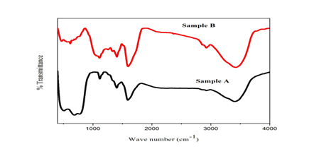
Figure 2a shows the FTIR spectrum of TiO2 nanoparticles in which the peaks corresponding to 3400.24 cm-1, in the spectra are due to the stretching of H - bond of the O - H (Alcohol) group, the peaks corresponding to 2923.73 cm-1 and 2255.35 cm-1 were indicated as the functional group of C-H (alkane stretching and - C ≡ C – (alkynes, variable not present in symmetrical alkynes). The peaks observed at 1593.09 cm-1 and 1404 cm-1 corresponds to C = C (medium weak multiple bands). The obtained products are the result of the organic compounds like vitamins, enzymes, monosaccharide, polysaccharide and lignin’s present in the Cynodon dactylon leaf powder (sample B). In Figure 2b all the peaks are assigned with that of Figure 2a.That means that the leaf compounds were mixed with TiO2 nanoparticles and is clearly seen in Figure 2a. In sample A, the peaks corresponding to 511.34 cm-1 , 686.81 cm-1 and 773.81 cm-1 show the stretching and vibration modes of O – Ti – O.

Figure 2 (A) FTIR spectrum of TiO2 nanoparticles of (A) Sample A and (B) Sample B (Cynodon dactylon leaf powder) shown in top most column
Raman spectroscopy
According to factor group analysis anatase has six Raman active modes which are 144cm-1(Eg), 197cm-1 (Eg), 399cm-1(B1g), 513cm-1(A1g), 519cm-1(B1g) and 639cm-1. In this part of study, four active Raman modes of 145 cm-1 (Eg), 399 cm-1 (B1g), 516 cm-1(B1g) and 639 cm-1 (Eg) for anatase TiO2 are evaluated for the sample A. Compared to the reference sample, the intensities of sample A are decreased which confirm the absorption of the bio molecules of Cynodon dactylon leaf extract by the sample A surface Figure 3.
Scanning Electron Microscope
To analyses the morphological studies of nanoparticles, the Field Effect Scanning Electron Microscopy is used. Figure 4 shows FESEM image of TiO2 nanoparticles. In the Figure 4, it can be seen that, the particles agglomerate with each other. In the SEM analysis particles were found to be irregular shape.
Transmission Electron Microscope
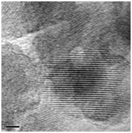
The morphology, crystallinity and size of the green synthesized TiO2 nanoparticles were also determined by TEM images. The shape of the nanoparticles was hexagonal and irregular in shape with moderate variation in size (Figure 5). The size was in the range of 13 – 34 nm. The average size of the nanoparticles was found to be 16 nm. The selected area electron diffraction pattern indicates, green synthesized TiO2 nanoparticles are in anatase phase with good crystallinity with dotted concentric rings which can be assigned to non spherical shape of TiO2, which is also assigned with XRD analysis. These are all shown in Figure 6.


Figure 5 N=57; Epidemiological distribution of the pathological fractures, traumatic fractures, and nonunion.
Antimicrobial activity
The synthesized TiO2 nanoparticles range from 10, 20, 30 and 40μm and they are using to determine the antibacterial effect by agar well diffusion method. The TiO2 showed (Figure 7) antimicrobial activity when tested against the pathogens. The antibacterial activity is found to be concentration dependent (i.e.), the antibacterial activity increased with the increase in the concentration of TiO2. The zone of minimum inhibition concentration was measured in a range of 15mm in 10μm. It is evident from the result that the cells were highly sensitive to all tested concentrations of the TiO2 nanoparticles, which was confirmed from the size of the zone of inhibition.

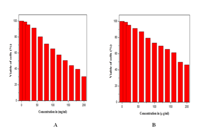
Figure 7 Anticancer activity for A549 cell lines for (A) commercially available TiO2 and (B) green synthesized TiO2 NPs.
The TiO2 nanoparticles exhibited a good antibacterial activity against Gram – negative bacteria (Table 1). The effect of antibacterial activity was minimum when the inhibition concentration was 10μm. This result is possible due to the difference in the concentration of TiO2 reacting in the gram negative cell wall. The cell wall contains a thinner layer of peptidoglycan. The TiO2 interacts with the membrane permeability of the bacteria and cleaves the cell wall resulting in the killing of bacteria. It is evident from the result of Figure 6 that the TiO2 nanoparticles possess potent bactericidal activity.
|
TiO2 Nanoparticles Concentration (μm) |
Diameter of Zone of Inhibition (mm) |
|
10 |
15 |
|
20 |
17 |
|
30 |
16 |
|
40 |
19 |
|
Control |
12 |
Table 1 Well Diffusion of TiO2 nanoparticles against E.coli
Anticancer activity (MTT assay)
Anticancer activity of A549 cell lines was studied using cell viability assay. IC50 value for A549 cell lines is 140 µg/ml. In 100% of live cells, 50% of cell is death when our synthesized nanoparticles were injected to the cells. That concentration value is IC50 value. In the above Figure 7 shows the commercially available TiO2 and green synthesized TiO2. Commercially available TiO2 IC50 value 200 µg/ml.16 In the above result, green synthesized TiO2 have good anticancer activity because in low concentration high cells are death. That means, bio molecules in Cynodon dactylon is giving excess electron to TiO2 super oxide radicals O2- are formed, the super oxide radicals produces ROS (Reactve Oxygen Species) in cancer cell lines. That ROS is used to break the cancer wall. Super oxide radicals production increased, ROS production is also increased. These are all reasons for electron production from TiO2. So leaf extract is working for high amount of electron production. So our green synthesized TiO2 nanoparticles efficiency is high for anticancer activity (A549 cell lines).
The TiO2 nanoparticles were synthesized successfully with Cynodon dactylon leaf extract by green synthesis method. The physico - chemical properties of synthesized TiO2 nanoparticles were investigated by XRD, FE-SEM, HRTEM and Raman spectroscopy analysis indicating the properties of synthesized TiO2 nanoparticles. Titanium dioxide nano particles show the inhibitory effect on the growth of E.coli, and enhanced anticancer activity against A549 (lung cancer) it was confirmed by the above said parameters.
None.
None.
None.

©2017 Hariharan, et al. This is an open access article distributed under the terms of the, which permits unrestricted use, distribution, and build upon your work non-commercially.