Journal of
eISSN: 2373-4396


Mini Review Volume 5 Issue 3
School of Medicine, Commemorative Hospital, USA
Correspondence: Breijo-Marquez Francisco R, East Boston Hospital, 02136 Tremont St., Boston, Massachusetts, USA
Received: February 07, 2016 | Published: February 18, 2016
Citation: Francisco BMR. A breijo pattern associated to a wolff-parkinson-white pattern. J Cardiol Curr Res. 2016;5(3):119-122. DOI: 10.15406/jccr.2016.05.00161
Increasingly is better known for different cardiologists the ‘Breijo pattern’ on an ECG tracing, characterized by the presence of a short PR (or PQ) interval and a short QT interval within the same ECG. Wolff-Parkinson-White syndrome is much better known and studied. The Association of both patterns on the same person can, considerably increase the symptomatology in the patient and also can make this symptomatology much more serious, even endanger the patient’s life. In the paper, the author has wanted to show to interested readers this association with a young woman of 26 years-old with mild but frequent symptoms. She had also had seizures, tonic-clones that were treated incorrectly. To emphasize that she had also had some of syncope events by undiagnosed cause.
Careful assessment of an ECG tracing is prescriptive when a patient reports symptoms suggestive of cardiac alterations, because in many cases such ECG tracings may go unnoticed. The results can be fatal if they are not diagnosed.
Keywords: cardiac arrhythmias, tachyarritmias, syncope, sudden cardiac death, breijo pattern, wolff-parkinson-white pattern, delta wave, short PR- interval, short QTc interval, rare cardiac disease
In 2008, Breijo-Márquez1 published an article in the “International Journal of Cardiology”, whose content was the description of an ECG pattern unknown to date. This pattern consisted of a shortening of PR and QT- intervals in the same individual (on an ECG tracing). The article was entitled by the author as “Decrease of electrical cardiac systole”. Since then, another many cases with same ECG features have been assessed and diagnosed.
At many times this pattern is present with other different cardiac pathologies. Thus, for example, Breijo-Marquez et al. have found this “Breijo Pattern” associated with a “Wellens syndrome “(also know as Wellens pattern).2 Breijo-Márquez et al. have also published a case with the presence of a Wolff - Parkinson-White pattern associated with a long QT interval in an intermittent way.3
In order for such ECG pattern can be considered as a “Breijo pattern” should fulfill the following features in the ECG tracing: the length of both intervals must be in these ranges:
Breijo pattern is only such if a shortened PR and QTc intervals are present on the same ECG tracing. Just in this case!.1–5
There are many formulas for calculating the corrected QT-interval, depending on heart rate, measured by the length of the RR- interval (in milliseconds) (Table 1). But the most frequently used by most cardiologists are still the Bazett and Fridericia formulas, and we usually make use of these formulas for calculating such measures.
|
QT heart rate correction formulas |
|
|
Exponential |
Formula |
|
Bazett |
QT/RR1/2 |
|
Fridericia |
QT/RR1/3 |
|
Linear |
Formula |
|
Framingham |
QT+0.154 (1-RR) |
|
Hodges |
QT+1.75 (HR-60) |
Table 1 Formulas for QTc measure
While all authors agree that the length of the PR-interval should be equal or greater than 120 milliseconds and equal or lesser than 200 milliseconds, when it comes to agree on what must be the outcome for a QT- interval for be considered as shortened, there are many disagreements among authors. The same happens when a QT- interval should be considered as Long: there is no unanimity among the various authors on when it should be considered as such. They are so many the discrepancies among the different authors that, for some of them, it cannot be considered as a “short” QTc -interval if its length does not is lesser than 320 milliseconds (Depending on their own convenience, possibly). For us, the corrected QT-interval (Bazett and Fridericia’s correction) must have a value higher than 350 milliseconds for to be considered as within normal limits. Regardless of the age and sex of assessed person.
So much so that, we think that all symptoms- by milder that they can be- and can make us think in the cardiac sphere, should be deeply investigated, using as a “fundamental weapon” for its diagnosis a 12 lead electrocardiogram surface.
Breijo pattern features1–5
In the Breijo pattern, the most significant feature is the presence of a short PR -interval and a QTc- interval in the same ECG of the patient.1 The symptoms can be imprecise-or very significant, endangering the life of the patient by the sudden appearance of tachyarrhythmias - ventricular normally - that require fast acting, because otherwise an unexpected death may occur. The symptoms may be isolated or in mixtures; they don’t have to appear all symptoms at the same time. However, in front of any suggestive symptoms, we must perform a 12 lead ECG and make a profuse assessment there of. The most frequent symptoms are:
Fortunately, the most common symptoms are also the most milder. However a cardiac arrest may also appear debuting at this event, which can also be very difficult to recover, especially if it happens in a place not specialized.
Wolff-parkinson-white pattern features6–9
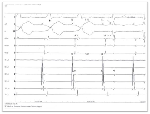
It is not our intention to make this document an exhaustive exhibition of the different types of Wolff-Parkinson-White Pattern that have been described and are already well known; just we want show to interested readers the existence of a Wolff-Parkinson-White Pattern and a Breijo pattern in the same ECG tracing of a 26 year- old patient with symptoms suggestive of cardiac alteration. As is already well known, Wolff-Parkinson-White Pattern, can be very clear or, on the other hand, can appear intermittently on an ECG tracing. In this exposed case, the delta wave is very subtle as well as “intermittent”. Nevertheless, the EEP test was positive for WPW (Figure 1).10

Figure 1 EEP image: Long anterograde refractory period. Stimulation of the distal coronary sinus a S1S1 cycle of 580ms with maximum preexcitation and sudden the accessory pathway anterograde conduction block. An antegrade refractory period and anterograde conduction 1:1 over the accessory bundle can be seen.10
Basically, Wolff-Parkinson-White pattern6 is characterized by the association of an anomaly in the cardiac electrical conduction (accessory pathway) and the occurrence of arrhythmias. It is a syndrome of pre-excitation of the ventricles of the heart due to an accessory pathway known as the bundle of Kent that runs across the part anterolateral from the right atrium and right ventricle (if by the left known as bundle of Ohnell).
The Kent bundle is an accessory pathway conduction between the cardiac atria and the ventricles. It is an abnormal path that is present in a small percentage of the general population. This is a bundle of tissue which can be found either between the left atrium and the left ventricle, whose case is called type A pre-excitation, or between the right atrium and the right ventricle, in whose case is called type B. The preexcitation troubles arise when this pathway creates an electrical circuit that avoids the AV node. The AV node has the property of reducing the speed of the electrical impulses, while the path that is set through the bundle of Kent not. When performing an abnormal electrical connection through this bundle, tachyarrhythmia events can occur.
Patients are usually asymptomatic. However, during episodes of supraventricular tachycardia, the individual may experience palpitations (perception of the own heartbeat), dizziness, difficulty breathing, and sometimes lightheadedness. We must never forget that the most typical image, nearly pathognomonic, of a Wolff-Parkinson-White pattern is none other than the “delta wave”.
In Wolff-Parkinson-White pattern, the most common symptoms are very similar to the Breijo pattern.
An ECG tracing with the WPW pattern will show the characteristic “delta wave”.
However, it should be noted that the delta wave, so characteristic of Wolff-Parkinson-White Pattern, can appear and disappear in the same ECG tracing. It is the called “intermittent delta wave”.3,6,7
According to some clinical studies, the “intermittency” of the delta wave in the ECG tracing, presupposes a major probability that an individual with such “intermittency” can remain asymptomatic throughout life. Individuals with Wolff-Parkinson-White pattern in which delta waves disappear with increasing heart rate are the ones with the lower risk of sudden cardiac death. This is because the loss of the delta wave shows that the accessory pathway cannot drive the electrical impulses at a high rate (in the anterograde direction). In general, these individuals will not have a quick driving by the accessory pathway during episodes of atrial fibrillation.
Although the vast majority of people with Wolff-Parkinson-White pattern remain asymptomatic throughout their lives, there is a risk of sudden death associated with this pattern. It is rare (occurring in less than 0.6%), and is due to tachyarrhythmias caused by this accessory pathway (Kent or Ohnell bundles) in the individual.
Illustrative clinical case
On this occasion, we will present the ECG of a 26-year-old adult woman who has suffered for a long time ago (from her childhood) several vague symptoms of intense palpitations-mainly at night and at rest -, sudden dizziness, a blurred vision as well a as a generalized profuse sweating. Also is worth to say that, during her early childhood, the patient had suffered several attacks of tonic-clonic seizures of a short duration and with a total recovery. Such seizures were classified as “epileptic seizures “, despite the fact that was never been seen any “focal point of epilepsy” in the different electroencephalograms that were performed on the patient. Also is worth to say that, during her early childhood. In our experience, it is quite common that the patients with a Breijo pattern or the WPW pattern begins with symptoms of seizures (tonic-clonic, mainly) which are treated as epilepsy in an empirical manner: In this clinical case, any epileptic focus was noted.
All these symptoms were self-limiting, so that when the woman went to several emergency Centers to be assessed, the symptoms had already disappeared or they were already very mild. However, on two occasions, she had suffered two events of a true syncope, contrasted and commented by their closest relatives; These events of syncope were not related to effort. The recovery of them was complete, without aftermath.
At several times, in Emergency rooms, the patient was undergoing to all kinds of medical studies (ECG, echocardiography, blood analysis - with included cardiac markers -) giving anodyne results, and without any security to give a definitive diagnosis: the patient just was stabilized of her symptoms by the physicians and she was discharged to her home after of her stabilization. Until she was finally diagnosed with this ECG association.
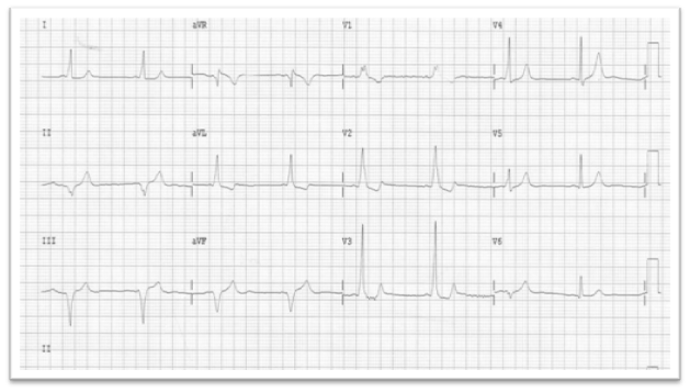
Once the patient was definitely diagnosed with these associated patterns, a conservative treatment was programmed (Propafenone, at a rate of 450 milligrams per day). Depending on developments with such treatment, it was proposed to the patient the possibility of a selective Ablation by radio frequency, after preceptive electrophysiological study (EEP). During two years, the patient has not had any symptoms like those taht she had been mentioned (before being diagnosed with these associated patterns). We have therefore preferred to maintain the conservative treatment, without forgetting the possibility of Ablation.After being braked the heart rate by administering of beta-blockers (Atenolol type) her ECG tracing can be seen in Figure 2.


Figure 2 ECG tracing from the 26-year-old woman taken at rest, at a heart rate between 50-51 beats per minute (slight bradycardia) after having been "braking" through the administration of Atenolol. Among other things, we can see the presence of a delta wave, mainly in D1 during the first heartbeat and in anteroseptal precordial leads: V4-V5, as well as a shortening of PR and QT intervals. We can see the last ECG tracing that was obtained after having slowed her heart rate up to 50 bpm, by means of Atenolol. We do not have images of ECG prior to this tracing. According to patient, these have been lost or cannot be found.
The measures of the different intervals give us shorter lengths for the PR and QTc-intervals on the same ECG tracing (Breijo pattern) (Figure 3).1 Namely, a Breijo pattern associated to a Wolff-Parkinson-White pattern on the same ECG tracing.1–5

Figure 3 Enlarged image from main ECG (in detail on V4-V5). Can be seen the presence of a Delta-wave.
The PR interval length is lesser than 120 milliseconds (short PR, intermittent delta wave). The QT length is lesser than 360 milliseconds (357 milliseconds), The RR-interval length is 1,224 milliseconds (50- beats per minute). The QTc length is lesser than 350 milliseconds:
That is to say, a typical “Breijo pattern” (Table 2).
|
RR |
1.224489795918 |
seg |
|
QTc (Rautaharju) |
440 |
mseg |
|
QTc (Bazett) |
323 |
mseg |
|
QTc (Framingham) |
357 |
mseg |
|
QTc (Fridericia) |
334 |
mseg |
|
QTc (Call) |
330 |
mseg |
Table 2 Breijo pattern
We are then in front of an ECG tracing, presenting:
That is, a Breijo pattern and a Wolff-Parkinson-White pattern together on the same ECG tracing of a person with mentioned symptoms previously.
Summing up, we ought to insist that all suggestive symptoms of cardiac disorder should be studied profusely. The exposed clinical case is not an anecdotal case, since we have seen these (or similar) cases in many other occasions. Facing a to event similar to exposed, all cardiologists ought to do an exhaustive measure of all segments and intervals from the ECG tracing in order of to make a correct diagnosis, and therefore to make a correct treatment. In this way, the events underdiagnosed, like the exposed here, would be much more diagnosed and thus, would have a more suitable treatment.11
None.
The authors declare there is no conflict of interests.
None.

©2016 Francisco. This is an open access article distributed under the terms of the, which permits unrestricted use, distribution, and build upon your work non-commercially.