Journal of
eISSN: 2373-4396


Case Report Volume 3 Issue 1
1Erebouni Cardiology Center, Yerevan, Armenia
2Klinik Im Park, Zurich, Switzerland
Correspondence: Hayrapetyan HG, Department of Urgent Cardiology, Erebouni Medical Center, Titogradyan street 14, 0087 Yerevan, Armenia, Tel 37491505005, Fax 37410473800
Received: May 19, 2015 | Published: July 18, 2015
Citation: Hayrapetyan HG, Gasparyan VC, Vogt PR. Giant left atrial diverticulum with thrombus inside: case report. J Cardiol Curr Res. 2015;3(1):1-6 DOI: 10.15406/jccr.2015.03.00091
A 59-year-old woman was admitted to the CCU due to atrial fibrillation and heart failure signs. Transthoracic echocardiography showed a huge intracavitary thrombus (7x8 cm) originating from left atrial diverticulum (7x8 cm). A coronary angiogram was performed and revealed substantial stenosis of diagonal brunch. The patient was treated by surgical removal of the diverticulum combined with an aortocoronary bypass of diagonal brunch. The patient has remained in sinus rhythm for 1 day after surgery. No complications were observed in postoperative period, and the patient was discharged on the 5th day following surgery.
Keywords:left atrium, diverticulum, thrombus
AF, atrial fibrillation; LA, left atrial; LV, left ventricle
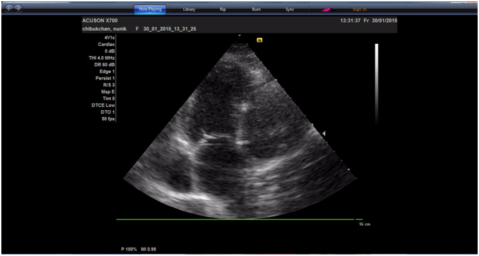
A 59 year-old female with a history of atrial fibrillation (AF), and arterial hypertension presented to CCU with respiratory insufficiency, palpitation and weakness. She had developed AF 2-3years earlier and was followed up at the outpatient clinic. She had been treated with warfarin at a dose that resulted in an international normalized ratio of 2.0 to 3.0. She was also taking beta-blocker, ACE inhibitor. Blood pressure at admission was 140/80 mm Hg, blood oxygenation was 90-92%. Electrocardiography showed atrial fibrillation with the ventricular rate of 90-100 per minute. Transthoracic echocardiography revealed a giant (7 x 8cm) left atrial (LA) diverticulum with big (5 x 6cm) thrombus inside (Figure 1). Systolic function of the left ventricle (LV) was preserved; LV ejection fraction was 50%. The diverticulum was compressing the left atrium and left ventricle lateral wall.

Figure 1 Transthoracic echocardiographic apical four-chamber view demonstrating the diverticulum interconnected with the left atrium.
CT-scan revealed big cavernous formation (7 x 8cm) draining into the LA with big oval mass (5 x 6cm) inside its cavity. The patient underwent coronaroangiography and the significant stenosis of diagonal brunch of LAD was observed. The patient underwent excision of the diverticulum and aorto-diagonal bypass grafting. Aortic and two-stage right atrial cannulation was performed and cardiopulmonary bypass was instituted with 32OC cooling followed by antegrade cold blood cardioplegic solution.
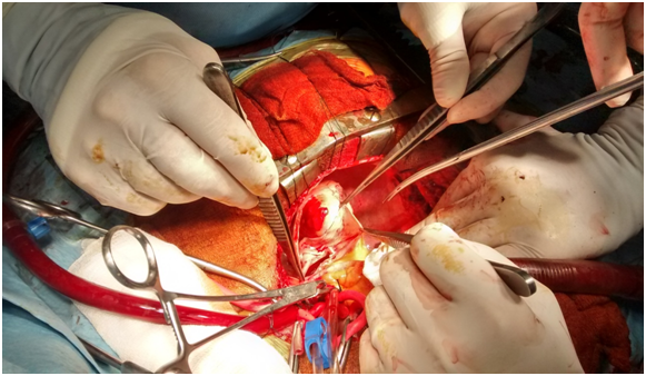
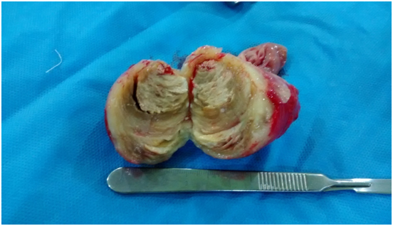
The diverticulum was excised and the thrombus from its cavity was sent for histology (Figure 2 & 3). LA appendage was sutured with continuous double layer Prolene 4/0 suture. No obvious cauliflower-like formation was found on the left atrium wall, and the diagnosis of thrombus as well as diverticulum was confirmed by histology. The normal sinus rhythm recovered on first postoperative day.

Figure 2 Intraoperative picture of the diverticulum excision. Giant left atrial diverticulum communicating with the atrial cavity is seen.

Figure 3 Big white organized thrombus removed from the diverticulum cavity with calcified nucleus(central zone).
The postoperative course was uneventful. All haemodynamic parameters were in normal range. The patient was discharged with normal sinus rhythm on fifth postoperative day. The patient has remained in sinus rhythm for 3months postoperatively, and transesophageal echocardiography revealed no LA thrombus, no evidence of residual LA diverticulum.
Accessory LA appendages and atrial diverticulum have an incidence of 10-27%.1 They are commonly found on cardiac-gated CT.2 But their association with atrial fibrillation needs to be confirmed. Some researchers reported that LA diverticulum could be found in 36% of patients with AF.3,4 A literature review reveals many clinical cases reporting LA diverticulum complicated with AF, yet our case is unique for huge sizes of both diverticulum and thrombus in it.
Cardiac diverticuli are rarely encountered. They are mostly asymptomatic and may be discovered incidentally. That is why the diagnosis is made much less than they exist truly. We describe the unusual case of a 59-year-old woman who was referred to our hospital with a giant left atrial diverticulum with thrombus formation and persistent atrial fibrillation. The mass of the diverticulum compressed the left atrium and left ventricle. The diverticulum was surgically excised and the thrombus removed. The normal sinus rhythm recovered spontaneously just after surgery and the the patient remained asymptomatic with very good quality of life during three months of follow up period.
None.
Author declares there are no conflicts of interest.
None.

©2015 Hayrapetyan, et al. This is an open access article distributed under the terms of the, which permits unrestricted use, distribution, and build upon your work non-commercially.