Journal of
eISSN: 2378-3184


Research Article Volume 8 Issue 1
Department of Marine Science Mawlamyine University Myanmar
Correspondence: Thet Htwe Aung Demonstrator Marine Science Department Mawlamyine University Myanmar
Received: November 23, 2018 | Published: January 24, 2019
Citation: Aung TH. Two species of marine brown algae (Phaeophyta) recorded in Kalegauk Island, Myanmar. J Aquac Mar Biol. 2019;8(1):7-8. DOI: 10.15406/jamb.2019.08.00234
The samples were collected from the different stations along the Kalegauk Island from August 2016 to January 2017. Based on the external morphology and internal structures, the samples were identified under the compound microscope and dissecting microscope. A total of two species of two genera were identified. The names of the species are Colpomenia sinuosa (Mertens ex Roth) Derbes̀ & Solier and Dictyota adnata Zarnardini. They all were new records for algae resources of Kalegauk Island and they were found in all stations abundantly. The present study is to record the diversity of marine brown algae and study their morphology from Kalegauk Island.
Keywords: Kalegauk, Colpomenia sinuosa, Dictyota adnata, morphology
Kalegauk Island is the island in Ye township, Mon state, Myanmar. It is located in the northern part of the Andaman Sea, 8.25km from the coast of Mon. The island has a long shape with a length of over 10km and a width of 1.6km in its widest area and there is a small Cavendish island lies 0.5km off the southern point of Kalegauk Island. In the study areas, salinity range and temperature regimes seawater were 26-27‰ and 29°C to 31°C, respectively. Nowadays, Kalegauk Island has been declared as the island for the future deep sea port.
Marine benthic green algae were collected from Apor Seik, Pashyu Chaung, Chaytoryar Pagoda, Alè Seik, Auk Seik and Kyunn Pyet from August 2016 to January 2017 (Figure 1). In the laboratory, the collected samples were identified on taxonomic characters. For internal studies of the thalli, cross sections were obtained manually with shaving blades. Vegetative and reproductive structures of the plants were studied under the Olympus compound microscope and Kaneko Yushima dissecting microscope. Microscopic measurements were recorded in micrometer (µm) using the ocular meter. The species were identified by using Zarnardini, Soe Pa Pa Kyaw, Pham et al., Silva, Bass on and Moe, Taylor, Dawson, Khin Khin Gyi, Kyaw Soe and Kyi Win.1-8
Microthalli yellowish-brown, globular, hemispherical, hollow, smooth, to irregularly convoluted, expanding to 1-2cm or more in diameter and filled with water when young but collapsed later. Plants are sessile and attached by a broad basal disc, saccate portion, membranous and crisp in texture. Cross section of the membrane showing cortex of 1-2 layers of small cuboidal or polygonal cells, about 8-28µm in diameter, chromatophores present. Medulla with 2 to 4 layers of colorless cells, irregularly in shape, 20-160µm long and 20-80µm wide (Figure 2A) (Figure 2B).
Dictyota adnata Zarnardini
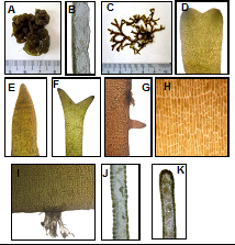
Thallus flattened, ribbon-like, brown to golden brown in color, prostrate with dentate margin, repeatedly about 1.5-2.5cm in height, repeatedly dichotomous branched, and attached by marginal rhizoidal processes scattered along the edges of the thallus. Filaments of rhizoid are 375-500µm long and issuing downward from the underside margin of thallus for secondary attachments. Branches are non-twisted with obtuse or sometimes acute apices. Secondary branches 350-500µm long and 150-170µm broad. Cells of the surface view elongated, arranged, 20-40µm long and 8-16µm wide. In cross section, thallus differentiated into a cortex and a medulla. Cortex cells two rows above and below margin of the thallus, small, rectangular and thin walled cells with many chromatophores, 8-16µm in diameter. Medullary cells one rows in the middle of thallus, large, rectangular and thick walled cells without chromatophores, 40-60µm long and 15-32µm wide (Figure 2C-2K).

Figure 2 Brown algae of Kalagauk I. (A) Habit of Coipomenia sinuosa (Mertens ex Roth) Derbes̀ and Solier. (B) Cross section of C. sinuosa (Mertens ex Roth) Derbes̀ and Solier. (C) Habit of Dictyota adnata Zarnardinii. (D-F) Apical portion of D. adnata Zarnardinii. (G) Branchlets of D. adnata Zarnardinii. (H) Rhizoidal filaments of D. adnata Zarnardinii. I) Surface view of D. adnata Zarnardinii. (J) Cross section of middle portion. (K) Cross section of upper portion.
Tin Aung Moe et al.,9 was firstly observed the seaweeds found around the Kalegauk Island in 1971. After that, some marine algae of Kalegauk Island had been described as a small part of their algal flora study in Myanmar by Kyaw Soe and Kyi Win8 in 1977. However, there were no brown species recorded in their studies. As a result, these two species can be regard as new records for Kalegauk Island. As for diversity of Myanmar algae resources, Colpomenia sinuosa and Dictyota adnata were recorded by Kyaw Soe and Kyi Win8 and Soe Htun.10 In addition, Khin Khin Gyi7 observed the morphology and distribution of Colpomenia sinuosa (Mertens Ex Roth) Derbes & Solier from Myanmar. Soe Pa Pa Kyaw2 had studied on the genus Dictyota of Myanmar which had based on a total of six species of the genus Dictyota. However, the two species, Colpomenia sinuosa and Dictyota adnata could be seen all around the Kalagauk Island significantly. In addition, Colpomenia sinuosa are growing on rocks or bottom surface in shallow water or tide pool, easily detached and cast ashore and it was found that Dictyota adnata grows close to high tide level mainly on sandy-silty mangrove.
None.
Author does not have any conflicts of interest.

©2019 Aung. This is an open access article distributed under the terms of the, which permits unrestricted use, distribution, and build upon your work non-commercially.