Journal of
eISSN: 2378-3184


Research Article Volume 12 Issue 2
1School of Marine Sciences, Pontifical Catholic University of Valparaiso, Chile
2School of Applied Marine Sciences, Universidad de Oriente, Venezuela
Correspondence: José Patti, Department of Aquaculture, School of Applied Marine Sciences, Universidad de Oriente, Venezuela
Received: May 25, 2023 | Published: July 31, 2023
Citation: Valencia Z, Patti J, Gaviria J. Embryonic development of the blenny Parablennius marmoreus (Osteichthyes: Blenniidae). J Aquac Mar Biol. 2023;12(2):188-193. DOI: 10.15406/jamb.2023.12.00373
The fish Parablennius marmoreus (Blenniidae) exhibits demersal spawning characterized by eggs adhering to solid surfaces and male parental care. This research describes the embryonic development under laboratory conditions of 12 spawns obtained in captivity at temperatures ranging from 27 ºC to 30 ºC. The first cell division (2 blastomeres) was observed at 50 minutes post-fertilization (PF). The eggs are smooth, light orange in color, with differentiated violet chromatophores. The chorion diameter measures 0.62±0.06 mm, and the granular yolk has a diameter of 0.45 ±0.08 mm. The blastula stage occurred at 11 hours and 40 minutes PF, and gastrula at 21 hours and 10 minutes PF. Embryo formation took place at 48 hours PF, and the larvae hatched at 135 hours PF, measuring 2.91±0.34 mm in length with a notochord and displaying 97 beats per minute. The captive reproduction and embryonic development of the blenny P. marmoreus are feasible and offer a solution to contribute to the conservation of species with ornamental interest.
Keywords: demersal spawning
Parablennius marmoreus inhabits very shallow bottoms, typically at depths of less than 10 m, on solid substrates with rocks and/or gravel, in areas with relatively clean and clear waters, although not necessarily coral formations. It is also found in seagrass meadows of Thalassia testudinum.1
The reproduction of this group of fish is characterized by the deposition of eggs that adhere to substrates such as empty mollusk shells and barnacles, as well as in rock crevices and dock pilings, which are used as nests where they are incubated, typically by the male, until the larvae hatch.2–4
Caribbean Sea in Venezuela, it is a common species and sometimes very abundant in certain areas, such as Charagato Bay in Cubagua. It is easy to observe as it always swims close to the substrate, where it spends most of its time at rest. It is also frequently found in areas with empty bivalve shells, where it seeks refuge, as well as in mussel and oyster cultivation rafts, among the shells and vegetation attached to the ropes. It exhibits various color variations, ranging from uniform dark brown to those with a prominent lateral stripe and rounded brownish-orange spots on a light background.1
The blenny Parablennius marmoreus is of great interest in aquaculture. Its appeal as an ornamental fish lies in the variety of colors it exhibits. According to Wabnitz et al.,5 based on data from the Global Marine Aquarium Database (GMAD) from 1997 to 2002, blennies accounted for 2% of the total trade of 24 million marine ornamental fish, comprising 10 major groups (Pomacentridae, Pomacanthidae, Acanthuridae, Labridae, Gobiidae, Chaetodontidae, Callionymidae, Microdesmidae, Serranidae, and Blenniidae).
Several descriptions of embryonic and larval development have been conducted for species within the Blenniidae family, including Hypsoblennius jenkinsi and H. gentilis,6 Lypophrys pholis,7 L. trigloides,8 Parablennius pilicornis,9 P. ruber,10 P. gattorugine, and P. ruber.11
Given the importance of Parablennius marmoreus in the field of aquaculture and the need to generate information regarding the embryonic development of this species, the present study aimed to describe the embryonic development of this species under laboratory conditions.
Study area
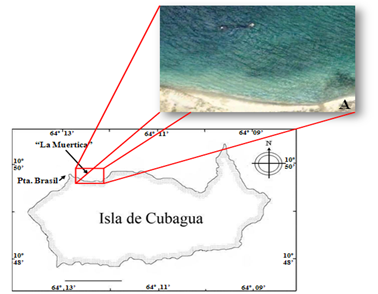
Cubagua Island (Figure 1), located 10 km from the town of Punta de Piedras (Margarita Island), was chosen as the breeding site. It is situated between 10º 47' and 10º 51' north latitude and between 64º 8' and 64º 14' west longitude in the Caribbean Sea. The island covers an area of 23 km2. Its coastline features rocky ecosystems, sandy beaches, seagrass meadows, and coral patches.12

Figure 1 Cubagua Island, Venezuela Caribbean (A) Sampling area "La Muertica" (10º 49'40" N and 64º 11'59" W), located near Punta Brasil lighthouse. Image taken from Google Earth.
Data collection
In the northwestern part of the island, in an area called "La Muertica" (10º 49'40" N and 64º 11'59" W), near Punta Brasil lighthouse, a semi-sunken vessel approximately 20 m long was found about 90 m from the shoreline. The fish were manually collected using dip nets at a depth of approximately 1m.
Pair selection and spawning
In the breeding room of the Institute of Scientific Research (IIC) at UDONE, Venezuela, a circular tank with 600 liters of filtered seawater was used to house the blennies for a period of two weeks for acclimation and pair formation.
Once the pairs were formed, they were removed and each pair was placed in white fiberglass circular tanks with a capacity of 50 liters (3 tanks), filled with 20 liters of filtered seawater. Two PVC pipes of ½ and 1 inch were placed in each tank, where the fish deposited their eggs. The PVC pipes were cut in half and joined together with a rubber band, allowing the viable and non-viable eggs to be counted and identified once the fish spawned inside the pipe.
Fecundity
When spawns were found in the PVC pipes in the circular tanks, the pipe containing the egg mass was immediately removed and replaced. It was then placed in a small aquarium (12 liters capacity), where the eggs were incubated until hatching.
Upon placing the pipe in the aquarium, the number of spawned eggs was counted, and viable and non-viable eggs were identified. The eggs were differentiated based on their coloration, with fertilized eggs being violet in color and unfertilized eggs appearing pale yellow, almost transparent.
A sample of viable eggs was then collected from the PVC pipe using a small spatula. The eggs were placed in a Petri dish with a certain amount of water from the same aquarium and transported to the IIC's ichthyology laboratory for further observations.
Embryonic development
In the ichthyology laboratory of the IIC, samples of the eggs spawned by the Parablennius marmoreus breeders were observed. An Olympus BH2 microscope was used to describe the stages of embryonic development according to the criteria established by Nikolsky in 1963,13 following the methodology described by Wittenrich14 for the development of blenny species belonging to the Meiacanthus genus.
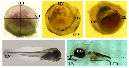
Photographs were taken during embryonic development using a MOTIC MOTICAM 2300 digital camera with a resolution of 3.0 megapixels, adapted to the aforementioned microscope. The camera's digital program was used to calibrate the equipment with the microscope's objectives (following the instructions in the camera's manual) in order to measure the following characteristics, as established by Ortiz et al.,15 egg width (AH), egg height (HH), vitellus diameter (DV), vitellus height (HV), perivitelline space (EPV), oil droplet diameter (DGA), notochord length (LN), post-orbital distance (DPO), rostrum length (LR), eye diameter (DO), gut tube length (LTD), mouth length (LB). The appearance and development of optic and otic capsules, heart, Kupffer's vesicle, pectoral fins, and pigment cells were also observed (Figure 2).

Figure 2 Morphometry of eggs and larvae of Parablennius marmoreus, indicating the points from which measurements were taken. AH, egg width; HH, egg height; DV, vitellus diameter; HV, vitellus height; EPV, perivitelline space; LN, notochord length; EE, embryo axis; DPO, post-orbital distance; LR, rostrum length; DO, eye diameter; LTD, gut tube length; LB, mouth length.
The volume of the vitellus (VV) and oil droplet (VGA) during the stages of embryonic development was calculated using the formulas proposed by Heming and Buddington in 1988:16
V = 0,1667 π L H2 (1)
V = 0,1667 π D3 (2)
Where,
L: length of the yolk
H: height of the yolk
D: diameter of the oil droplet
Feeding of the fish
The breeders were fed ad libitum three times a day, every day, in the morning and evening with balanced marine fish feed, and at noon with frozen mysids. Excess food that the fish did not consume was removed by siphoning the tank bottom with a small hose to prevent bacterial proliferation in the tank.
Physicochemical parameters
During the experiment, variables such as salinity were recorded using a Milwaukee MR100ATC refractometer with an accuracy of±1 ppt, while temperature and dissolved oxygen of the water were measured using a YSI 550A model with an accuracy of±0.1 ºC and±0.01 mg/l, respectively.
Data processing
Averages and standard deviations of egg and larval measurements were calculated using spreadsheet software, specifically Microsoft Office Excel 2007.
Description of spawning
The breeding pairs spawned in the ½ and 1-inch PVC pipes, usually in the morning hours. The eggs were consistently placed in the middle part of the pipe's interior.
Continuous spawns were recorded in 3 pairs of P. marmoreus, totaling 9,074 eggs obtained in captivity from a total of 12 spawns, with a range of 134 to 2,315 eggs per spawn, occurring from July to November (Figure 3).
Description of the eggs
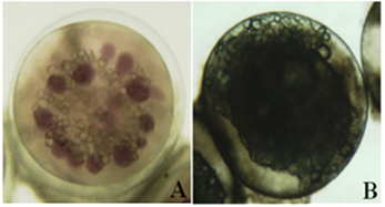
The eggs of P. marmoreus are elliptical, smooth without ornamentation, and slightly flattened on the side that adheres to the substrate. Fertilized eggs displayed a light orange color, with differentiated violet chromatophores in the yolk (Figure 4A). The hue of the eggs intensified as the embryo developed, turning into a deep brown or bright golden shade when the larva was about to hatch. The yolk is spherical, granular, and lacks oil droplets. The perivitelline space is clearly distinguishable. Unfertilized eggs exhibited a pale yellow, almost transparent coloration with disorganized yolk (Figure 4B).

Figure 4 Spawned eggs of Parablennius marmoreus. A) Fertilized egg without division B) Unfertilized egg.
Description of embryonic development
Fertilized eggs without division (T=0) have an average width and height of 0.62±0.04 mm and 0.47±0.01 mm, respectively, and exhibited a light violet coloration due to the dispersed chromatophores in the yolk. The yolk is granular with an average diameter of 0.52±0.01 mm and a volume of 0.08 mm3. The perivitelline space is transparent with an average width of 0.11±0.04 mm (N=6). No lipid droplets were observed (Figure 4A).
The first division (formation of the two initial cells) was observed at 0 hours and 50 minutes post-fertilization (PF). The animal pole was differentiated. The egg coloration remained the same as in the previous stage (Figure 5). The average diameter of the yolk was 0.52±0.04 mm. The average volume of the yolk was 0.08 mm3. The average width of the perivitelline space was 0.11±0.04 mm (N=3).
The formation of the 32 blastomeres was observed at 6 hours post-fertilization (PF) (Figure 6). The average width and height of the egg were 0.62±0.02 mm and 0.47±0.01 mm, respectively. The average diameter and volume of the yolk were 0.45±0.02 mm and 0.06 mm3, respectively. The average width of the perivitelline space was 0.18±0.01 mm (N=4).
The morula stage, following the formation of 32 cells, was identified. Clear cell aggregation (blastomeres) was observed at the animal pole. At 11 hours and 40 minutes post-fertilization (PF), the blastula stage was observed, which appeared as a hollow sphere larger than the morula, with the blastocoel inside. The average width and height of the egg were 0.62±0.03 mm and 0.47±0.01 mm, respectively. The yolk had reduced in size, with an average diameter of 0.44±0.04 mm and an average volume of 0.05 mm3. The average width of the perivitelline space was found to be wider (0.2±0.04 mm). The average diameter of the oil droplets (N=13) was 0.2±0.1 mm, with an average volume of 0.10 mm3 (N=54). During the blastula stage, the invagination process began, forming a cavity inside and initiating gastrulation.
The gastrula stage (Figure 7) was observed at 21 hours and 10 minutes PF. The yolk was bilobed and exhibited small oil droplets that merged as development progressed. The blastodisc or germ ring was visible. The embryonic shield was observed in several eggs, indicating the initiation of embryo formation. The average width and height of the egg were 0.62±0.04 mm and 0.47±0.01 mm, respectively. The average diameter of the yolk was 0.42±0.06 mm, with an average volume of 0.045 mm3, indicating a reduction in size. The average width of the perivitelline space was 0.2±0.05 mm, which increased as development progressed and was later occupied by the embryo. The average diameter and volume of the oil droplets were 0.5±0.3 mm and 0.24 mm3, respectively (N=45).

Figure 7 Egg of Parablennius marmoreus in the gastrula stage: embryonic shield, oil droplets in the bilobed yolk.
The neurula stage (embryo formation) was observed at 48 hours and 10 minutes post-fertilization (PF). As the embryo developed, different structures could be observed, including the Kupffer's vesicle, optic capsules, protocerebrum, notochord, pectoral fins, embryonic fin, heart, myoseptum, optic capsules, remnants of the digestive tube, and chromatophores (Figure 8 & 9). The average width and height of the egg were 0.62±0.07 mm and 0.47±0.02 mm, respectively. The average diameter of the yolk was 0.38±0.1 mm, with an average volume of 0.02 mm3, indicating a reduction in size. The average notochord length ranged from 0.83±0.16 mm in the early stage to 1.51±0.36 mm in the late stage. The average embryo axis was 0.33±0.17 mm, the average post-orbital distance was 0.57±0.16 mm, the average rostral length was 0.10±0.05 mm, and the average eye diameter was 0.15±0.04 mm (N=97). At 79 hours PF, the remnants of the pectoral fins were observed, and the embryo exhibited 76 beats per minute (BPM) and mobility within the egg. At 119 hours and 30 minutes PF, the formed ocular lenses were observed, and 97 BPM were counted.

Figure 8 Developing embryo of Parablennius marmoreus in the early stage: optic capsules, chromatophores.

Figure 9 Developing embryo of Parablennius marmoreus in the advanced stage: eyes, remnants of pectoral fins.
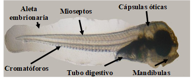
The hatching of the larvae (Figure 10) was observed at 139 hours post-fertilization (PF) after the complete absorption of the yolk. The average notochord length was 2.91±0.34 mm. Upon hatching, the larva exhibited 97 BPM, a two-chambered heart, well-developed eyes (0.28±0.03 mm) with fully formed lenses, and a golden coloration. The mouth was fully open (0.66±0.12 mm) and ready for exogenous feeding. The average rostral length was 0.10±0.05 mm. The optic capsules were observed with their two pairs of otoliths, sagittae, and lapilli (sacular and utricular).

Figure 10 Larva of Parablennius marmoreus: Mandibles, optic capsules, myosepta, embryonic fin, chromatophores, and digestive tube.
The average post-orbital length was 0.87±0.23 mm, and the average length of the digestive tube was 0.88±0.19 mm. The larva exhibited a large number of fully visible myosepta, with 30 chromatophores along the notochord. Body fluids were visible throughout the body, from the heart to the caudal region of the larva, and vice versa. The embryonic fin was present and visible from the dorsal region of the head to the ventral region where the anus is located. The pectoral fins were well-developed. The larva exhibited active swimming behavior (N=5).
Physicochemical variables
The salinity was maintained at 37 ups throughout the 5-month duration of the experiment. The dissolved oxygen level averaged 5.7±0.03 mg/L. The average temperature was 28.7±1.2 ºC, ranging from a recorded minimum of 25.6 ºC to a recorded maximum of 31.6 ºC.
Number of eggs (fecundity)
In this study, a total of 9,074 eggs were obtained in captivity from P. marmoreus, with an average of 12 spawnings and an interval of 134 to 2,315 eggs per spawning. Stevens and Moser6 reported that a female Hypsoblennius jenkinsi, 141 days after hatching, spawned 450 eggs in the tank where it was kept. This female continued spawning for 6 months, with an interval of 300 to 650 eggs per spawning, but none of these eggs survived. Locatello and Neat17 reported that female Aidablennius sphynx laid between 253 and 402 eggs per spawning, depending on the size of the males they chose as their partners for reproduction. The embryonic and larval description of Lipophrys polis7 and L. trigloides8 was based on 3 spawnings, while for Parablennius pilicornis, it was based on 2 spawnings, all obtained in captivity. However, the number of eggs per spawning was not recorded in these studies.9
Description of eggs
Hempel18 mentioned that different characteristics of fish eggs can help differentiate them, especially during simultaneous spawnings, such as egg shape and size, chorion structures, yolk pigmentation, and the number and size of oil droplets. The freshly spawned fertilized eggs of P. marmoreus described in this study are similar to those reported for Blennius pholis, B. gattorugine, and B. ocellaris (AH = 1.4 mm, 1.6 mm, and 1.2 mm, respectively, Lebour19), Hypsoblennius jenkinsi (AH = 0.75±0.029 mm, Stevens and Moser6), Lypophrys pholis, L. trigloides, Parablennius pilicornis, P. gattorugine, and P. ruber (AH = 1.3±0.04 mm, 1.3±0.16 mm, 0.60±0.07 mm, 1.2±0.05 mm, and 1.1±0.05 mm, Faria et al.,7–9,11), P. marmoreus, Meiacanthus grammistes, M. nigrolineatus, and Petriscirtes breviceps (AH = 1.0 mm, Wittenrich,14), P. ruber (AH = 1.02±0.03 mm, Villegas et al.,10), and Salaria fluviatilis (AH = 1.2±0.05 mm, Gil et al.,20).
Description of embryonic development
At 79 h PF in P. marmoreus eggs, the embryo exhibited 76 BPM, and at 119h: 30 min PF, the ocular lenses, otoliths, digestive tube, and pectoral fins were formed, the heart had two chambers, and 97 BPM were counted. Faria et al.,7 reported that during the embryonic development of Lipophrys pholis, pigmented eyes, brain, notochord, myomeres, heart, digestive tube, otic capsules, and developed pectoral fins were observed at 216h PF. In Parablennius gattorugine and P. ruber, similar characteristics were described at the same stage of development,11 as well as for the description of Lipophrys trigloides at 240 h PF.8 In Parablennius pilicornis, these characteristics were described at 164 h PF,9 and in Salaria fluviatilis, they were described at 264 h PF.20
The hatching of P. marmoreus larvae was observed at 139 h PF (LN =2.91±0.34 mm; 25.6-31.6 ºC). Upon hatching, the larvae exhibited a large number of visible myosepta, with 30 chromatophores along the notochord. Faria et al.7 reported that the larva of L. pholis hatched at 360 h PF (LT = 5.03±0.19 mm; 15.5-17.5 ºC). The larva of L. trigloides hatched at 432h PF (14 and 16.5 ºC) and 312h PF (21 and 23 ºC), with a total length of 4.8±0.19 mm.8 In Parablennius pilicornis, larval hatching occurred at 216 h PF (LT = 3.1±0.07 mm; 18-20 ºC), and these larvae can be distinguished from other representatives of the same genus because they have 6 or 7 preopercular spines.9
Wittenrich14 stated that P. marmoreus larvae hatched approximately at 240h PF (LT = 3.2 mm), with immediate exogenous feeding. In Meicanthus grammistes, hatching occurred between 168 and 192h PF (LT = 3.0 mm; 26.6 ºC), in M. nigrolineatus between 192 and 240 h PF (LT = 3.7 mm; 24.4-26.6 ºC), and in Petriscirtes breviceps at 168 h PF (LT = 3.0 mm; 27.2 ºC). In P. gattorugine, larval hatching occurred at 340 h PF (LT = 5.2±0.07 mm; 17.5 and 19.5 ºC), and in P. ruber at 384 h PF (LT = 4.1±0.07 mm; 16 ºC). Both larvae can be differentiated because P. ruber has 2 to 4 preopercular spines, while they are absent in P. gattorugine.11 Villegas et al.,10 reported that newly hatched (still with yolk) P. ruber larvae measured 4.65±0.25 mm in total length. Gil et al.,20 stated that the larva of the freshwater blenny, Salaria fluviatilis, hatched at 312 h PF (LT = 5.1±0.07 mm; 20-21 ºC).
Ajith et al.,21 mentioned that the trade of ornamental marine fish is rapidly growing, and it is mostly based on the extraction of these animals from their natural habitat. More than 1,000 species of coral reef fish are collected for the ornamental trade. The increasing demand for ornamental marine fish can lead to the depletion of wild populations. Cultivating coral reef species could alleviate the growing market demands and help increase wild populations, as well as generate employment opportunities.
The captive breeding of the blenny species P. Marmoreus under controlled conditions does not seem to be a difficult task, which benefits aquaculturists when it comes to mass production of this group of fish. Therefore, these results indicate a positive response and a solution to contribute to the conservation of these species and thus reduce the extraction from their natural habitat for ornamental purposes.
None.
The authors declare that there are no conflicts of interest.

©2023 Valencia, et al. This is an open access article distributed under the terms of the, which permits unrestricted use, distribution, and build upon your work non-commercially.