eISSN: 2373-6372


Case Report Volume 5 Issue 1
1Department of Pathology, Apollo Gleneagles Hospital, Kolkata, India
2Institute of Gastrosciences, Apollo Gleneagles Hospital, Kolkata, India
Correspondence: Amrita Chakrabarti, Department of Pathology, Apollo Gleneagles Hospital, Kolkata, India
Received: May 28, 2016 | Published: July 7, 2016
Citation: Chakrabarti A, Goenka MK (2016) Submucosal Lipoma of the Rectum Presenting with Rectal Prolapse and Appearing as an Adenomatous Polyp on Confocal Laser Endomicroscopy: a Case Report and Review of Literature. Gastroenterol Hepatol Open Access 5(1): 00126. DOI: 10.15406/ghoa.2016.05.00126
Lipomas of the gastrointestinal (GI) tract are rare neoplasms, with rectum being the most uncommon site of occurrence. Most of these lesions are asymptomatic; however, the larger sized ones may have varied presentations ranging from vague abdominal discomfort to over symptoms simulating malignancy. Prolapse of the rectum is a very rare presentation of rectal lipomas and warrants further investigations to avoid misdiagnosis. Colonoscopy and endoscopic ultrasound (EUS) are important modalities for preoperative diagnosis; nevertheless, which remains difficult in some cases. Confocal laser endomicroscopy (CLE) is a novel endoscopic imaging modality that allows tissue diagnosis at cellular and sub cellular levels, and bears particular significance to the diagnosis of polypoidal lesions. However, application of CLE for sub mucosal lesions such as lipomas of the colorectum has not being documented yet to the best of our knowledge. Here we report a case of lipoma of the rectum presenting with features of rectal prolapse. Colonoscopy and CLE were performed, the findings of both of which were suggestive of an adenomatous polyp, thereby providing no diagnostic advantage of CLE over white light endoscopy for this particular case of rectal lipoma. However, EUS revealed a sub mucosal lesion. Subsequently, sub mucosal endoscopic dissection (SMD) of the mass was performed, that was later confirmed to be a sub mucosal lipoma histopathologically. This reiterates the importance of histopathology for the diagnosis of GI lesions over novel “virtual microscopy” techniques.
Keywords,sub-mucosal rectal lipoma, rectal prolapsed, confocal laser endomicroscopy, adenomatous polyp
EUS, endoscopic ultrasound; SMD, submucosal endoscopic dissection; CLE, confocal laser endomicroscopy; GIST, gastrointestinal stromal lesion
Lipomas of the colon and rectum are rare GI neoplasms, though they are the second most common benign tumors after adenomatous polyps,1 and the commonest non-epithelial neoplasms. They comprise about 5% of all GI tract tumors,2 with an incidence ranging from 0.035% to 4.4%.3 The most frequent sites are the ascending colon, ceacum and sigmoid colon.4 Rectal lipomas are even rarer, constituting only 5% of the total GI lipomas.5 About 90% cases arise from the sub mucosa, with the remaining 10% being subserosal or intramuscular.6 Most colorectal lipomas are asymptomatic and incidentally detected during colonoscopy, surgery or autopsy. However, those greater than 2cm may induce symptoms such as bleeding, obstruction, intussusception, anemia, abdominal pain, a change in bowel habit and rarely prolapsed.7
Preoperative diagnosis is usually missed, with most being presumed to be adenomas or carcinoma.8,9 GI lipomas become clinically significant as they are likely to be confused with carcinomas, especially in symptomatic lesions, resulting in extensive surgeries that may result in higher rates of mortality and morbidity.10 As a result, preoperative diagnosis remains challenging. Imaging modalities include barium X ray, ultra-sonography (USG), computed tomographic (CT) scan, magnetic resonance imaging (MRI) and EUS, however, the findings are generally non specific. Colonoscopic appearance is that of yellowish polyps that may be sessile or pedunculated, with intact overlying mucosa, which may rarely be necrotic or ulcerated.
The past few years have witnessed the emergence of new endoscopic imaging modalities for the diagnosis of GI lesions. CLE is a novel technology allowing surface histological diagnosis at a cellular and sub cellular level in vivo, therefore having the potential to provide instantaneous optical diagnosis for neoplastic polyps and replacing histopathology.11 However, definitive diagnosis can only be made by histopathological evaluation in post resected specimens.6 Symptomatic lesions less than 2cm are usually removed by endoscopic resection, whereas larger tumors stand a risk of perforation and should be removed surgically by open or laparoscopic methods.12 However, endoscopic removal is preferable over surgical excision, provided the cases are selected carefully and the procedure performed by skilled endoscopists in centers of excellence.1
We report a case of rectal lipoma in a 61year old female presenting with symptoms of rectal prolapse. Investigations included white light endoscopy, CLE and EUS. Colonoscopic examination revealed a 4cm sessile polypoidal growth. This was followed by CLE, which showed complete disorganization and loss of glandular architecture, consistent with an adenomatous polyp. EUS was suggestive of a gastrointestinal stromal lesion (GIST) or a lipoma. SMD of the lesion was subsequently performed, and was confirmed to be a lipoma histopathologically.
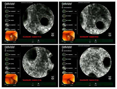
A 61years old lady presented to the Gastroenterology outpatient department with a one month history of vague lower abdominal pain, altered bowel habit and the sensation of something passing through the anus during defecation, which she manually reinserted. There was no associated tenesmus, bleeding or weight loss. She was then admitted and investigated further. Colonoscopy was performed, which revealed a polyploidal growth about 4cm in size with smooth surface, which was about 1cm from the anal verge (Figure 1). This was followed by CLE, the findings of which included a complete disorganization and loss of glandular architecture in the involved area with diffuse inflammatory cell infiltration (Figure 2). Our centre is the first one in India to use CLE technology for diagnosing GI lesions. Moreover, this is the first case where CLE has being used for a GI lipoma, to the best of our knowledge.

Figure 2 Confocal laser Endo microscopy showing complete disorganization and loss of glandular architecture with diffuse inflammatory cell infiltration.
The colonoscopy and CLE findings were consistent with an adenomatous appearing polyploidal mass of the rectum. Thereafter, EUS was performed, which showed a 5.7x20 mm homogenous, hyper echoic lesion arising from and limited to the second layer, or, the muscularis mucosa of rectum, with normal surrounding vascularity and absence of lymph nodes. The findings were suggestive of a gastrointestinal stromal tumor (GIST) or a lipoma. Subsequently, she underwent endoscopic mucosal dissection of the mass, (Figure 3) which was sent to histopathology as a rectal polyp.
Grossly, the specimen was a single intact mucosa covered polypoidal piece of tissue measuring 4x3.5x2cm in size. Histopathological analysis revealed a submucosal tumor comprising of lobules of mature adipose tissue with intervening fibrous septa, confirming a lipoma (Figure 4). There was no dysplasia or malignancy. Her remaining course in the hospital was uneventful and was eventually discharged with the advice to follow up within a month.
Lipomas of the GI tract occur most frequently in the right colon, comprising 70% of all cases. In the descending order of prevalence, the other locations include the transverse colon, descending colon, sigmoid colon with rectum being the rarest site of occurrence. They have also being reported in the small intestine, stomach and esophagus.12 These lesions have been found to occur most frequently in the 5th to 6th decade, with a slight female preponderance. The size usually ranges from 0.5 to 10cm.
Although lipomas are the second commonest benign neoplasm of the colon, their overall incidence is infrequent. A meta-analysis performed by Weinberg and Felman demonstrated their incidence to be only 0.2%,13 though the range varies from 0.035% to 4.4%. Lipomas of the rectum have been very infrequently reported; with extensive literature review revealing only 9 such cases being reported till date.14 Moreover, most of these lesions remain asymptomatic, only to be discovered incidentally during a colonoscopy, surgery or autopsy. Less than one-fourth of colonic lipomas have been reported to be symptomatic.12 However, lesions larger than 2cm may present in varied ways including abdominal pain, changes in bowel habit, intussusception, weight loss, anemia, GI bleed and prolapse. Such manifestations may raise clinical suspicion of a more ominous diagnosis.
Colorectal lipomas are of mesenchymal origin, arising from the submucosa and protruding into the bowel lumen. The etiology has not been clearly understood, although chronic inflammation may contribute to the development of these lesions due to abnormal intestinal motility, resulting in separation of the mucosa from the deeper submucosa and subsequent adipose tissue deposition in the separated tissue space.12
Lipomas of the rectum presenting with symptoms of prolapse has rarely been reported in literature.15 Moreover, definitive preoperative diagnosis of these lesions is often difficult to make, as they can be easily misdiagnosed as mucosal prolapse. The typical features in colonoscopy include a yellowish smooth submucosal lesion exhibiting “cushion sign” or “pillow sign” due to surface indentation on pushing with biopsy forceps, and also the “naked fat” sign that reveals underlying fat after a biopsy. EUS usually demonstrates a hyperechoic mass arising from the rectal wall.16 However, preoperative diagnosis is often difficult, with most lesions being diagnosed during laparotomy and definitively by histopathology. Moreover, these lesions are often misdiagnosed to be adenomas or carcinomas on white light endoscopy, especially in presence of surface ulceration or necrotic areas.12
CLE is a novel method for diagnosing GI lesions that provides real time high resolution imaging at approximately thousand fold magnifications with a resolution of about 1 micron, which enables visualization of mucosa and lamina propria as well as individual cells. Often termed as “virtual biopsy”, they simulate images seen in traditional histology. CLE is particularly useful for diagnosing polyploidal lesions of the colorectum. The expected main advantage of CLE over white light endoscopy is that of non reliance on histology, to cut down undesirable costs and avoid removal of lesions with no or little malignant potential.17 However, CLE appearances in cases of colorectal lipomas have not been documented till date. In this case, the confocal microscopic appearance was that of an adenomatous polyp with complete disorganization and loss of glandular architecture, hence providing no added advantage over white light endoscopy in the diagnosis. The definitive diagnosis was only made by histopathology subsequently. This reiterates the importance of histopathology despite development of novel endoscopic imaging modalities.
Treatment is usually done for symptomatic lesions over 2cm. Endoscopic resection is preferable over surgical removal, and is mostly done for pedunculated lesions less than 2cms in size, since larger or sessile lesions carry the risk of perforation. However, recent advances in endoscopic techniques allow consideration of endoscopic removal as an alternative to surgery for larger lesions.16 SMD allows en bloc resection of GI neoplasm and bears the advantage of enabling removal of large neoplasm, and at the same time, providing control over the size and shape of resection.18
Submucosal lipomas of the rectum are rare neoplasms that may present atypically as a rectal prolapse. Colonoscopy and EUS are important modalities for preoperative diagnosis of these lesions. CLE is a recently developed novel endoscopic imaging technique having added advantages over white light endoscopy and aims to replace traditional histopathology for diagnosis of GI lesions, bearing particular significance for assessment of polypoidal lesions. However, histopathology remains the gold standard for diagnosis of GI neoplasm, especially submucosal lipomatous lesions that may not be detected accurately by CLE.
None.
Author declares there are no conflicts of interest.
None.

©2016 Chakrabarti, et al. This is an open access article distributed under the terms of the, which permits unrestricted use, distribution, and build upon your work non-commercially.