eISSN: 2373-6372


Case Report Volume 4 Issue 3
Department of Gastroenterology, Max Superspeciality hospital, India
Correspondence: Ankur Gupta, Department of Gastroenterology, Max Superspeciality hospital, Mussoorie Diversion Road, Malsi, Dehradun, Uttarakhand, 248003, India, Tel 91-9450274612, 91-9634628271
Received: November 22, 2015 | Published: February 26, 2016
Citation: Gupta A (2016) Unusual Behaviour of Hepatocellular Carcinoma in a Patient with Liver Cirrhosis. Gastroenterol Hepatol Open Access4(3): 00099. DOI: 10.15406/ghoa.2016.04.00099
Hepatocellular carcinoma (HCC) is a feared complication of cirrhosis with poor outcome if untreated. Patients often present with worsening of previously known or unknown liver disease. Here we report a patient with liver cirrhosis with untreated HCC and remained well after a year of follow up with improvement in liver function and decrease in size of tumour.
Keywords: hepatocellular carcinoma, cirrhosis, malignant tumor, gastrointestinal bleed, jaundice, abdominal ultrasound, splenomegaly, ascites, upper gastrointestinal tract endoscopy
Hepatocellular carcinoma (HCC) is the most common type of liver cancer. It is a feared complication of cirrhosis. The outcome is usually poor especially in those who are diagnosed with advanced disease. Spontaneous regression of a malignant tumor is an extremely rare phenomenon and its incidence is estimated to be one per 60,000-100,000 cancer patients.1 We report a patient with liver cirrhosis with untreated HCC and remained well after a year of follow up with improvement in liver function and decrease in size of the tumour.
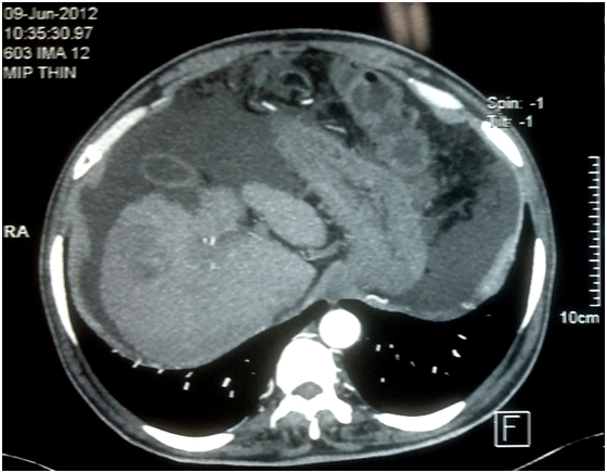
A 62 year male presented to our outpatient department with progressive abdominal distension for 1 month. He denied having fever, abdominal pain, jaundice, gastrointestinal bleed or encephalopathy. He did not smoke, consume alcohol, took any other drug or had any other illness. He lost 5Kg body weight during this illness. Investigations at presentation revealed haemoglobin of 12.3gm/dl (normal:13.5-17), total leucocyte count 9500/μL (normal: 4,000-10,000), platelet count of 152,000/μL (normal:150,000-400,000), prothrombin time 17 sec (control 13.1), creatinine 1.1mg/dL (normal: 0.8-1.4), albumin 2.6g/dL (normal: 3.5-5.4), bilirubin 1.8 mg/dL (normal: 0.3-1.5) and serum alpha-fetoprotein 4.4 ng/ml. MELD score was 13. Etiologic workup for cirrhosis was unrevealing. Abdominal ultrasound showed a shrunken nodular liver with a space occupying lesion in the right lobe of liver, dilated portal vein, splenomegaly and ascites. Upper gastrointestinal tract endoscopy showed small esophageal varices. Dynamic computed tomography scan of abdomen showed irregular atrophic liver with a rounded lesion of size 5.6X4.8 cm in segment V enhancement in the arterial phase (Figure 1) and washout in the portal-venous phase (Figure 2). Performance status was 3 on ECOG scale.

Figure 1 Arterial phase of computed tomography scan of abdomen showed irregular atrophic liver with an enhancing rounded lesion of size 5.6 X 4.8 cm in segment V.
Patient declined specific treatment for HCC and was treated with diuretics (furosemide 20 mg and spironolactone 50 mg daily). Ascites responded well and subsequently resolved completely. Diuretic dose was reduced to half after 10 months and his performance status rated on ECOG scale improved to 1. His investigations after 11 months of first presentation showed creatinine of 0.99 mg/dL, albumin 3.6 g/dL, bilirubin 1.5 mg/dL, prothrombin time 15.6 sec (control 11.8) and alfa-fetoprotein was 24 ng/ml. MELD was 12. An ultrasound abdomen showed the lesion size as 5.1 cm X 3.6 cm, portal vein diameter of 13mm, spleen size15.8 cm, no ascites, and no portal vein thrombosis.
We have described a patient with cryptogenic liver cirrhosis who presented with HCC (as evidenced by typical findings on computed tomography scan of the abdomen) and ascites. With his poor performance status at presentation, he had terminal stage HCC according to Barcelona-Clinic Liver Cancer classification system. After a year of follow up he was found to have a spontaneous reduction in tumour size, improvement in liver function and performance status.
Spontaneous resolution of HCC has been rarely described. Oquiñena & co-workers2 described partial or complete spontaneous regression of HCC in 3 patients and reviewed literature of 65 such patients reported earlier. The causative factors and mechanisms leading to spontaneous regression of HCC are unclear, however two causes have been considered.3 Firstly ischemia due to multiple causes like disruption of the feeding artery,4,5 portal vein tumor thrombus,6 massive bleeding, and poor arterial supply in cirrhotic liver causes necro-sis of the oxygen-sensitive malignancy and subsequent regression. Secondly immunological mechanisms might cause spontaneous regression of HCC.7 The mechanism of spontaneous regression in this case could not be ascertained. We conclude that spontaneous resolution of HCC is a rare phenomenon. Although awareness of such occurrence is important for hepatologist, its rarity should not prevent them from offering the standard treatment to patients with HCC.
None.
Author declare that there is no conflict of interest.
None.

©2016 Gupta. This is an open access article distributed under the terms of the, which permits unrestricted use, distribution, and build upon your work non-commercially.