eISSN: 2373-6372


Research Article Volume 3 Issue 2
Department of Gastroenterology, Hospital Universitario de Caracas, Venezuela
Correspondence: Daniel Tepedino Peluso, C.I. V15.646.405. Av. Humboldt cruce con Calle Baldo Urb. Bello Monte, Residencia Crystal Plaza, piso 4, Apto 4-B, Parroquia El Recreo, Municipio Libertador, Código postal 1050, Caracas-Venezuela, Tel 5.84127071462
Received: October 23, 2015 | Published: December 4, 2015
Citation: Peluso DT, Torrealba CDJB, Useche HMP (2015) Upper Gastrointestinal Bleeding in Caracas Hemorragia Digestiva Superior en Caracas. Gastroenterol Hepatol Open Access 3(2): 00074. DOI: 10.15406/ghoa.2015.03.00074
Materials and methods: The aim of this research is to describe endoscopic findings in patients with acute Upper Gastrointestinal bleeding. The sample consisted of 131 patients with signs or symptoms of Upper Gastrointestinal Bleeding during the first half of 2012. Non-experimental, descriptive and prospective research. Mean, mode and median for numerical variables and percentages were used.
Results and discussion: During the first half of 2012 were performed 2602 endoscopies, a total of 136 endoscopies were maid whose indication was upper digestive bleeding. Endoscopy was performed on 131 patients, 52.2% were female, 36.8% of endoscopies were performed in people of 60year or older. 48.9 % of patients had no comorbidity. 80.1 % of patients received no medication. The most frequent endoscopic finding was Peptic Ulcer Disease (44.1%). In 70.6% of cases no endoscopic therapy was applied.
Conclusions: Study findings are similar to those described in the national and international literature, differences were observed by increased frequency of neoplasms, increased use of argon plasma coagulation and low recurrence of bleeding. This research extended several recommendations.
Keywords: upper gastrointestinal bleeding, hematemesis, melena, endoscopic findings, endoscopic treatment
With more than 10% mortality Gastrointestinal Bleeding is a relevant entity, those patients who survive an episode have 15% chance of having a second episode despite adequate medical and endoscopic treatment.About peptic Ulcer Disease, currently most common cause of upper gastrointestinal bleeding, it was established that has a great economic impact; in the US it is estimated that the cost of absenteeism and medical and endoscopic therapy associated could reach 5.65billion per year.1 A Dutch study estimated that the cost of the complications of peptic Ulcer disease: bleeding, perforation or the combination of both was for 2004 of €12,000, €19,000 and €26,000 respectively.2
Recent research reports that endoscopic injection of adrenaline alone is a inferior treatment, a second treatment modality should be used in most cases.The combination treatment approach has been shown to significantly reduce recurrent bleeding, need for surgery, use of blood products, length of hospital stay and mortality.3
The frequency of Gastrointestinal Bleeding, morbidity, mortality and costs associated with it in Latin American countries are less documented.This research aims to provide information that will improve the knowledge about Upper Gastrointestinal Bleeding in Caracas, Venezuela and Latin America, will collaborate with the optimization of human and material resources in gastroenterology services, emergency services and Digestive Endoscopy Units.
It was a non-experimental, descriptive and prospective research.The study aimed to describe the findings and therapeutic on Upper Gastrointestinal Endoscopy in patients with Upper Gastrointestinal Bleeding at the Gastroenterology Department of the Hospital Universitariode Caracas in 2012. The population consisted of patients evaluated by the doctors of the Gastroenterology Department of the Hospital Universitariode Caracas at the Emergency Room, hospitalization or consults during the first half of 2012 and that merited emergency endoscopic evaluation.The sample consisted of 131 patients over 12years showed unmistakable signs or symptoms of Acute Upper Gastrointestinal Bleeding and who underwent upper gastrointestinal endoscopy at the Department of Gastroenterology at that hospital. Variables like Age, Sex, First endoscopy or successively, comorbidities, use of anti-inflammatory drugs, use of Esteroids drugs, Endoscopic Findings and Endoscopic Therapeutic (if performed) were considered.
The Gastroscopies practiced were performed by doctors Gastroenterologists (sometimes the Researchers) or residents in training, under the supervision of Gastroenterologists belonging to the Department of Gastroenterology of the Hospital Universitariode Caracas. Measures of central tendency (mean, mode and median) for numeric variables and frequency (percentages for nominal) were used to report the frequency of endoscopic findings in patients with Upper Gastrointestinal Bleeding. Finally, the similarities and differences observed with other populations evaluated in other national and international hospitals were established.A statistical consultant oversaw the calculations in SPSS and other related procedures.
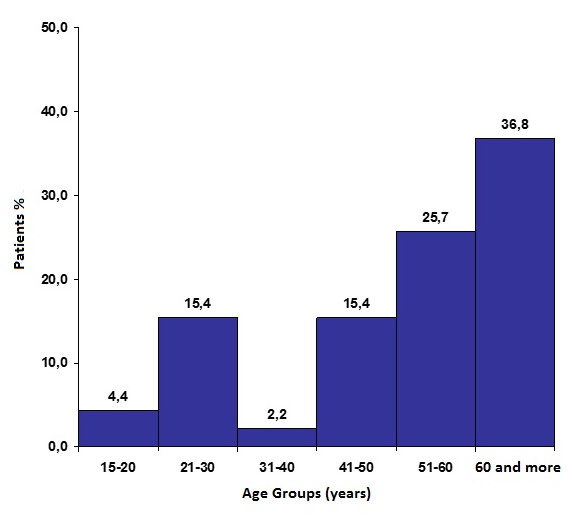
During the first half of 2012, 2602 digestive endoscopies were performed at the Gastroenterology Department of the Hospital Universitariode Caracas, 1565 were Gastroscopies and 1037 were Colonoscopies. A total of 136 endoscopies were performed in patients with Upper Gastrointestinal Bleeding. Gastroscopies were performed on 131 patients, 52.2% were female, ages ranged from 17-87 years (mean 53.6; mode 52, median 54, standard deviation ± 18.13 years).36.8% of Gastroscopies were performed in people 60 and older (Figure 1).

Figure 1 Endoscopic findings in patients with Upper Gastrointestinal Bleeding. Sample distribution by age.
No medical conditions were observed at 48.9% of patients, in the remaining 67 patients one or more conditions were observed within these the most frequent was High Blood Pressure (17.6%) followed by Ischemic Heart Disease and Liver Cirrhosis.Were observed several medical conditions: chronic, metabolic, vascular, renal, neoplasms and other diseases, and one pregnant. The 80.1% of patients received no medication, 18.4% were treated with NSAIDs and steroid 1.5%. The number of endoscopic findings varied from one (52.9%) to none (8.8%). The most frequent endoscopic finding was peptic Ulcer disease in 44.1% of cases (Table 1) placed more frequently at the Stomach (25%). The most common location of gastric ulcer was Antrum (74%) in the duodenal bulb in (51%). Forrest III Ulcers were observed in most cases (76.6% ofgastric and 59.8% of duodenal), followed by Forrest IIC (Table 2). Erosive Gastropathy (23.5%), Esophagitis (14.7%) Esophageal varices (13.2%) and Gastrointestinal Neoplasms (13.2%) were frequent findings.
Hallazgos endoscópicos |
n |
% |
Gastric Ulcer |
34 |
25.0 |
Gastric Erosions |
32 |
23.5 |
Duodenal Ulcer |
26 |
19.1 |
Esophagitis |
20 |
14.7 |
Esophageal Varices |
18 |
13.2 |
Portal Hypertensive Gastropathy |
16 |
11.8 |
Duodenal Erosions |
15 |
11.0 |
Gastric Cancer |
15 |
11.0 |
Hemorrhagic Gastropathy |
10 |
7.4 |
Mallory-Weisslesion |
5 |
3.7 |
Angiodysplasia |
5 |
3.7 |
Congestive Gastropathy |
4 |
2.9 |
Duodenal Tumor |
3 |
2.2 |
Gastroesophageal Varices tipo I |
2 |
1.5 |
Esophageal Foreign Body |
1 |
0.7 |
Normal |
12 |
8.8 |
Table 1 Endoscopic findings in patients with Upper Gastrointestinal Bleeding. Distribution of the sample according to endoscopic findings
70.4% of cases no endoscopic therapy was applied for not being indicated, 17.8% one method was applied and the remaining 11.8% a combination of two methods were used. The most commonly used monotherapy was the ligation of Esophageal Varices with elastic band (9.6%), it was applied to 13 patients who had Large Esophageal Varices (BAVENO) with active bleeding or stigmata of recent bleeding. The second monotherapy was Argon Plasma Coagulation (8.1%). The combined therapy more used was sclerosis with Adrenaline and later sclerosis with Alcohol (8.9%). Two cases required surgical treatment because of endoscopic therapy failure (1.5%, a patient with advanced gastric cancer and one with a pancreatic tumor with extension into the duodenum) (Table 3 & 4).
Ulcer |
n |
% |
Gastric |
||
Forrest III |
36.0 |
42.9 |
Forrest IIC |
5.0 |
6.0 |
Forrest IB |
4.0 |
4.8 |
Forrest IIA |
2.0 |
2.4 |
Duodenal |
||
Forrest III |
22.0 |
26.2 |
Forrest IIC |
7.0 |
8.3 |
Forrest IIB |
5.0 |
6.0 |
Forrest IB |
2.0 |
2.4 |
Forrest IA |
1.0 |
1.2 |
Total |
84 |
100.0 |
Table 2 Endoscopic findings in patients with upper gastrointestinal bleeding. Distribution of the sample according to location of the ulcer
Endoscopic therapy |
N |
% |
None |
95 |
70.4 |
Elastic Band Ligation |
13 |
9.6 |
Combined: Adrenaline and Alcohol Injection |
12 |
8.9 |
Argon Plasma Coagulation |
11 |
8.1 |
Emergency Surgery due to Endoscopic Therapy Failure |
2 |
1.5 |
Combined: Adrenaline injection and Argon Plasma Coagulation |
2 |
1.5 |
Total |
135* |
100 |
Table 3 Endoscopic findings in patients with upper gastrointestinal bleeding. Distribution of the sample according to endoscopic therapy
*A Esophageal Foreign Body was removed from one Patient but it was not considered therapy for Upper Gastrointestinal Bleeding.
Endoscopic finding |
Elastic band ligation |
Adrenaline and alcohol injection |
Argon plasma coagulation |
Adrenaline injection plus argon plasma coagulation |
Emergency surgery |
Total |
Large Esophageal Varices (BAVENO) |
13 |
13 |
||||
Advanced Gastric Cancer (Borrmann III) |
5 |
1 |
6 |
|||
Duodenal Ulcer (Forrest IIB) |
5 |
5 |
||||
Gastric Angiodysplasia |
5 |
5 |
||||
Gastric Ulcer (Forrest IB) |
4 |
4 |
||||
Gastric Ulcer (Forrest IIA) |
1 |
1 |
2 |
|||
Duodenal Ulcer (Forrest IB) |
2 |
2 |
||||
Duodenal Ulcer (Forrest IA) |
1 |
1 |
||||
Pancreatic Tumor with duodenal extension |
1 |
1 |
||||
Advanced Gastric Cancer (Borrmann IV) |
1 |
1 |
||||
Total |
13 |
12 |
11 |
2 |
2 |
40 |
Table 4 Endoscopic findings in patients with Upper Gastrointestinal Bleeding. Sample distribution by finding and therapeutics
Five patients required a second endoscopic examination because of recurrent or persistent bleeding (3.7% of cases): a man of 56years old with Gastric Cancer who required emergency surgery, a male of 68 years old with Pancreatic cancer duodenal extension required emergency surgery, a male of 42years with Liver Cirrhosis with a second Esophageal variceal Bleeding that was treated with another elastic band, a 31year old female with Stomach Ulcers without new endoscopic findings, a male of 48years old with Diabetes Mellitus type 2 had Gastric Angiodysplasia previously treated with Argon plasma without new findings (Colonoscopy revealed Angiodysplasia at Cecum and was treated with Argon Plasma coagulation). During the first half of 2012 Upper Gastrointestinal Bleeding was the indication 5.2% of all endoscopies and 8.7% of the Gastroscopies, being relevant.4
In terms of demographic characteristics, associated medical conditions and current or recent medication it was no difference with those described in the literature.5–9,10–12. Endoscopic findings were discordant with those published in America and Europe by Gastric Neoplasms (13.3%), Gastric Neoplasm were found to be more frequent than reported. This research describe one case of Esophageal Foreign Body as a cause of Upper Gastrointestinal Bleeding, a very rare cause.4,13 Analyzing endoscopic therapy used is noteworthy that in 70.6% of cases it was not required, suggesting the possibility that pre-endoscopic medical treatment administered was beneficial for patients with gastric ulcers and erosions, improving their prognosis, also it was associated with less severe cases of bleeding.4,6,14
Endoscopic Elastic Band Ligation and Argon Plasma Coagulation methods were widely used compared to that described in American literature, probably this was related to: 1) The high number of patients with Variceal Bleeding observed, 2) the fact that this is a Hospital with Hepatology Consult and postgraduate training and 4) because of the lack of such therapeutic methods in other Hospitals of the public health system.4,15–16
At the Gastroenterology department of the Hospital Universitariode Caracas combined methods for the control of Gastrointestinal Bleeding due to Gastric Ulcers were employed, it was observed the use of Adrenaline and Alcohol, and in conjunction with Argon Plasma Coagulation according to current recommendations of the American literature. Endoscopic injection of Adrenaline as monotherapy was no used.3,5,17
During the removing of aEsophageal Foreign Body (porkbone) which left a bleeding laceration that required Adrenaline and Hypertonic solution injection was documented, this is a rarecause of hematemesis and Upper Gastrointestinal Bleeding.4,13,18–19 The use of other endoscopic therapeutic methods such as Clips or Heater Probe was limited because of their availability. In accordance with current guidelines from the American Society for Gastrointestinal Endoscopy (ASGE), The Gastroenterology Department of the Hospital Universitariode Caracas showed lower mortality and recurrence of bleeding than described in American literature.4,5,14,20,21
This research contributes with information on the demographic and endoscopic characteristics of 131 patients who had Upper Gastrointestinal Bleeding during the first half of 2012 at the Hospital Universitariode Caracas. The most frequently involved was the female sex (52.2%), more than one third of patients were over 60years. 48.9% of patients showed no associated medical condition, the most common associated medical condition was High Blood Pressure (17.6%). 80.1% of patients received no medication, 18.4% used non steroid anti-inflammatory drugs and 1.5% steroids drugs.
The number of endoscopic findings varied from one (52.9%) to none (8.8%). The most frequent endoscopic finding was Peptic Ulcer disease in 44.1% of cases (Table 2) located more frequently at Stomach (25%). Erosive Gastropathy (23.5%), Esophagitis (14.7%) Esophageal Varices (13.2%) and Neoplasms Gastrointestinal (13.2%) were frequent findings. 70.4% of cases no endoscopic therapy was applied, 17.8% of cases one method was applied and the remaining 11.8% of cases a combination of two methods was used. The most commonly used monotherapy was the Elastic Band Ligation of Esophageal Varices (9.6%) followed by the use of Argon Plasma Coagulation (8.1%). The more used combined therapy was Adrenaline and Alcohol injection (8.1%). Two cases required surgical treatment because of Endoscopic Therapy Failure (Advanced Gastric Cancer and Pancreatic Tumor with duodenal extension). Five patients required a subsequent endoscopy because relapse or recurrence of bleeding (3.7% of cases).
Whereas the findings reveal that Peptic Ulcer disease is the most frequently cause of Upper Gastrointestinal Bleeding, themedical and paramedical training should focus on optimal management of this disease. Considering the large number of patients with variceal bleeding and the widespread use of Argon Plasma Coagulation, measures must be taken to ensure the availability of supplies and proper functioning of equipments.
The Hospital Universitariode Caracas lacks of intermediate care areas at both Emergency Room and Hospitalization, their creation will provide an opportunity to ensure optimal care to patients at high risk of death. Has been universally described the use of Risk Scores in patients with Gastrointestinal Bleeding, also the standardization of care with Gastrointestinal Bleeding protocols, their use could maximize resources and reduce associated morbidity and mortality.
Endoscopy records must be digitalized for ease of use, study and analysis. This records are an infinite source of valuable information that actually is very inaccessible and unfriendly to the user. Research should be promoted and encourage. Research and publishing must be a habit. The descriptions made in this research provide evidence of strong similarities between the evaluated sample and the Venezuelan and international population. These data should be complemented investigating causes of Lower Gastrointestinal Bleeding and Occult Gastrointestinal Bleeding and with the use of analytical statistics.
The authors want to thank all the staff of Gastroenterology Department of the Hospital Universitario de Caracas.
Author declares there are no conflicts of interest.
None.

©2015 Peluso, et al. This is an open access article distributed under the terms of the, which permits unrestricted use, distribution, and build upon your work non-commercially.