eISSN: 2378-3176


Case Report Volume 3 Issue 4
1Postgraduate department of urology, Chettinad health city, India
2Department of urology, Chettinad health city, India
Correspondence: Parikshith S, Postgraduate department of urology, Chettinad health city, Kelambakkam, Kanchipuram, Tamilnadu, India
Received: October 08, 2016 | Published: August 25, 2016
Citation: Parikshith S, Govindharaaju S (2016) Pelvi Neurofibroma Extrinsic Compression of Bladder. Urol Nephrol Open Access J 3(4): 00090. DOI: 10.15406/unoaj.2016.03.00090
Solitary neurofibroma occurs only in peripheral nerves. Pelvic Neurofibroma is one of the rare cases; we are reporting an encapsulated mass in the retroperitoneum on the right side causing extrinsic compression of the bladder.
Keywords: pelvic neurofibroma, extrinsic compression of bladder, solitary neurofibroma
Pelvic Neurofibroma is a very rare entity, so far approximately 60 cases reported.1 Usually Neurofibroma arise from the pelvic organs, but in this case tumor was not involved any pelvic organ.2 According to World Health Organization, it is an encapsulated nerve sheath tumor and common peripheral nerve tumors. About 10% are linked up with genetic neurofibromatosis type I (NF1). Neurofibroma without NF1 often described as slow growing, benign and painless tumor.3 In NF1 associated Neurofibroma are found with other diseases like skin discoloration, skin tags or small cutaneous tumor, Lisch nodules of the iris and central nervous system tumors such as acoustic neurilemoma or tumors arise from nerve roots of the spinal column. Patients with NF1 associated with 15% of malignant risk factors.4 Localized Neurofibroma with NFI usually seen in childhood and adolescence age. Multiple lesions usually seen along the deep and long nerve sheath.5
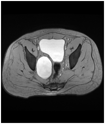
30 years old gentleman arrived with complaints of lower abdominal discomfort for 6 months with vague right iliac pain with normal gut and bladder habits. He experienced no other co-morbidities. Clinical examination findings were normal. Blood investigation was done within normal limits. Ultrasound abdomen showed a complex cyst with internal echoes and absence of vascularity in the right side of the pelvis. Contrast CT abdomen revealed non enhancing unilocular cystic mass on the right lateral pelvic wall.MRI, abdomen with gadolinium revealed T1 hypotenuse, T2/STIR heterointense enhancement within the lesion. Colonoscopy was normal. He underwent laparotomy under general anesthesia, through Pfannenstiel extra peritoneal approach mass lesion of size 8cm x 6 cm identified in the right pelvic region in the course of obturator nerve producing the compression over the lateral wall of the bladder without any infiltration of the bladder and bowel. It was arising from the nerve bundle, the lesion was well capsulated. It was highly vascular with blood supply from the branches of internal iliac arteries. The feeding arterial branch was ligated, outer capsule opened and an inner mass removed completely after leaving the capsule intact in view of nerve injury. Haemostasis achieved, postoperative period was uneventful. Histopathology revealed a mass of size 8x6.5x3.5cm section shows unencapsulated lesion comprised of spindle cells with wavy nuclei prominent myxoid stroma and scant collagen, conclusive of as Neurofibroma (Figures 1-4).

Figure 2&3 MRI, abdomen with gadolinium revealed T1 hypotenuse, T2/STIR heterointense enhancement within the lesion.
Neurofibroma is classified into two groups; first group is the solitary tumor or a non NF1 neurofibroma which is not associated with other tumors. Solitary Neurofibroma commonly occurs in female and occurs on the right side of the body. It occurs in later ages than NF1 associated Neurofibroma Second group of Neurofibroma is the plexiform Neurofibroma seen exclusively in conjunction with NF1. It has equal distribution in both the sides of the body and in sexes. In this study, Neurofibroma of pelvic origin producing extrinsic compression without any manifestation or involvement of Neurofibromatosis lesion. Only very less number of cases as has been reported without any malignant transformation. Narayana Pradeep et al.6 reported as a case of Neurofibroma involving phrenic nerve without any other neurofibromatosis lesion involvement.
Pelvic neurofibroma is the rare entity, usually associated with other organs; but unusual presentation may also present (not involving any pelvic organs). Surgical management is the only treatment available for compression symptoms or that Neurofibroma undergone malignant transformation.
Author declares that there is no conflicts of interest.

©2016 Parikshith, et al. This is an open access article distributed under the terms of the, which permits unrestricted use, distribution, and build upon your work non-commercially.