eISSN: 2377-4304


Case Report Volume 9 Issue 6
1Department of Obstetrics and Gynaecology, Simpson Centre for Reproductive Health, Edinburgh Royal Infirmary, UK
2Department of Pathology, Edinburgh Royal Infirmary& Western General Hospital, UK
Correspondence: Dr. Ibrahim Alsharaydeh, MRCOG, Specialty Registrar, Department of Obstetrics and Gynaecology, Simpson Centre for Reproductive Health, 51 Little France Crescent, EH 16 4SA Edinburgh, UK
Received: March 17, 2017 | Published: November 15, 2018
Citation: Alsharaydeh I, Jameel N, Oniscu A, et al. Vaginitis emphysematosa in 80 years old woman. Obstet Gynecol Int J. 2018;9(6):392-393. DOI: 10.15406/ogij.2018.09.00372
Introduction: Vaginitis Emphysematosa is a very rare condition, self limited cystic disorder of the vagina. It is caused by Trichomonos vaginilis and it is very common in pregnant or immunocomprimised patients. It can present with a pressure symptoms, abnormal cervical smear or vaginal discharge. In this paper, we present a case of this rare condition in an elderly lady.
Case report: An 80 years old woman presented to us with vaginal mass. She has had occasional smelly vaginal discharge, sensation of lump inside her vagina, with a sensation of not being able to completely emptying her bowels of 6 months duration. Local examination revealed a well circumscribed mid line cystic mass in the lower third of her vagina, measuring 2cm in diameter (size of a small lemon). Histological assessment of the biopsy revealed overall appearances are those of vaginitis emphysematosa.
Conclusion: This rare case would help clinicians and pathologists to be aware about the recognition of this unusual condition, which is a benign self limiting condition.
An 80 years old, nulli-parous woman. Who is generally fit and well. She was referred by her GP to the Gynaecological oncology rapid access clinic due to presence of vaginal mass with suspicion of vaginal malignancy. She has had occasional smelly vaginal discharge, sensation of lump inside her vagina, with a sensation of not being able to completely emptying her bowels of 6 months duration. Apart from chronic constipation, she denied any urinary, bowel symptoms with no vaginal bleeding. She had a total hysterectomy with bilateral salpingo-oophorectomy for large ovarian cyst at age of 47.1−4
On general examination, she was of an average build and her general health is good for her age. Local examination revealed a well circumscribed mid line cystic mass in the lower third of her vagina, measuring 2cm in diameter (size of a small lemon). There was white vaginal discharge with atrophic vulva and vagina. Also she had mild cysto-rectocele with no erythema, discharge and no other abnormalities were visualized. The regional lymph nodes were not enlarged. Digital rectal examination revealed solid hard stool with no diverticulum or fistula. Systemic examination revealed no abnormality.
Her routine blood tests; FBC, U&Es, VDRL and urinalysis were within normal limits. Punch biopsy taken from the centre of the mass and sent for histological assessment, a high vaginal swap sent and MRI was requested for further assessment of this cystic mass. High vaginal swap revealed presence of "clue cells" on microscopy, indicate infection with bacterial vaginosis. Her MRI didn’t reveal any other abnormalities.
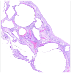
Histological assessment of the biopsy revealed sections show non dysplastic squamous epithelium with numerous variably sized micro cystic spaces. These changes were seen throughout the full thickness of the biopsy includes the basal cell layers of the epidermis.5−8
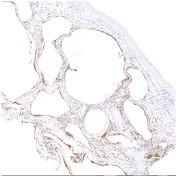
The smaller superficial spaces were denuded of any lining epithelium, while the larger ones within the stroma were partially lined by macrophages and foreign body giant cells. The features are very unusual, but suggestive of air spaces. Immuno-stains show negative epithelial markers and only CD68 positive macrophages lining the spaces. The overall appearances are those of vaginitis emphysematosa (Figure 1).


Figure 1 Histology reveals cystic lesions of variable size and foreign body-type giant cells in the cyst's wall, associated with minimal chronic inflammatory infiltrate. CD68 highlights the histiocytes and giant cells surrounding these cysts.
Patient was reassured about the findings. She was offered surgery to excise this cystic mass, but patient declined surgery due to her age and as the cystic mass not causing her any significant symptoms. The fore, patient was treated with one course of Metronidazol and discharged to her general practitioner.
The term “vaginitis emphysematosa” was coined by Zweifel in 1877. Vaginitis emphysematosa is a rare, benign condition and it is seldom diagnosed on examination as it lacks specific features to raise the clinical suspicion. Vaginitis emphysematosa is characterized by gas-filled cysts in the vaginal wall, in a pattern similar to pneumatosis of the intestines or bladder. This bacterial vaginitis is benign and self-limited and does not signify the presence of tissue necrosis or life-threatening infection.
Although the cause of this disorder is not completely understood, most cases have been associated with vaginitis caused by T. Vaginalis. Other organisms associated with this condition include Haemophilus vaginalis and Gardenerella vaginalis. Immunosuppression as a predisposing factor leading to the development of vaginitis emphysematosa has also been suggested.
Literature review showed that vaginitis emphysematosa occurs in a wide age range after menarche to well beyond menopause. Most patients, including our patient, have symptoms of vaginitis, including pruritis and a vaginal discharge. Occasionally patients complain of vaginal fullness and a bloody discharge. Symptoms are often short-lived and self-limited. The natural course for vaginitis emphysematosa is to resolve spontaneously and in most patients this has been an incidental Finding. On those occasions when the mass makes the patients seek advice simple excision may be done. Apart from solid tumors like condylomata acuminata, other benign cystic lesions of the vagina like inclusion cyst, Gartner duct cysts, and endometriosis should be considered in the differential diagnosis.9−11
Emphysematous changes in the ovary have not been described previously, has been well described in the literature. A case of emphysematous changes in the ovary of a 38-year-old woman; who presented with a right ovarian cyst. Histologically, a portion of the ovary had numerous cystic spaces lined by histiocytes and giant cells resembling those seen in Vaginitis emphysematosa. In addition, a case of Vaginitis emphysematosa which has predominantly affected the cervix has been reported. This has caused an abnormal cervical smear and Colposcopic investigation. In 1997, a cervico-vaginitis emphysematous is a rare self-limiting disease in which multiple gas-filled cysts are present in the submucosa of the upper vagina and ectocervix. A case report of a 40 year-old lady who presented with clinical features suggestive of carcinoma of the cervix. This indicates that the clinicians and pathologists have to be aware about the recognition of this unusual condition, which is a benign self limiting condition.
None.
The authors declare that they have no conflict of interest.

©2018 Alsharaydeh, et al. This is an open access article distributed under the terms of the, which permits unrestricted use, distribution, and build upon your work non-commercially.