eISSN: 2377-4304


The world has embraced minimal access surgery like no other modality due to the enormous benefits to the patients and surgeons. Faster recovery and early resumption of routine normal activities attract the population the most. Access into the abdomen is the foremost challenge of any laparoscopic surgery. The first entry into the abdomen is the most crucial and dangerous step in laparoscopic surgery as it is blind, thus amounting for complications like injuries to major vessels and gastrointestinal tract. A large Multicentric prospective study from 72 hospitals of the Netherlands revealed that the intestinal injuries are major complications during laparoscopy that occur in 5.7/1,000 procedures. Approximately 70% of these are related to the primary port entry. The overall incidence of laparoscopic entry injuries is 3.3/1,000 with gastrointestinal damage occurring in 1.3/1,000 and major vessel injuries in 1.05/1,000.1 At least 50% of the major complications occur prior to commencement of the intended surgery.2–4 means they are related to first blind primary port.
As minimal access surgery is having an upper edge over laparotomy not only for simple cases, but also for larger masses and gynaecological malignancies. Many gifted surgeons are in elite class of removing very large myomas and uterus laparoscopically.5,6 Other complex laparoscopic entries are made in clinical situations of previous surgeries done either laparoscopic or via laparotomy. Still more complex situations are laparoscopic removal of big masses with previous surgeries, and dealing gynae malignancies.7 As there is liberalization of caesarean section as a route of child birth, the situation of co-existent previous surgery in clinical indications for gynae laparoscopy are faced very often, like TLH with multiple previous caesarean sections. Rise in infectious pathologies; in gynae patients give rise to more adhesions, tubal blocks and then infertility that necessitate a laparoscopy. With the clinical scenario of previous surgeries, big masses, infectious pathologies and complex scenarios of combinations of these, laparoscopic entry poses certain challenges. Another challenge is also on rise - Obesity ǃǃǃ As the global incidence of obesity rises, gynae endoscopist find more patients in the obese and morbidly obese groups.8–10
So, however accomplished endoscopic surgeon is first blind entry in afore mentioned clinical situations, alone or in combinations, pose a definite challenge. More ever unfortunately, 30–50% of the bowel injuries and 15–50% of the vascular injuries are not diagnosed at the time of injury.1 This delay has contributed to mortality rates of 3–30% for bowel and vascular injuries.11,12 As the clinical scenario is changing, we need to refine laparoscopic entry techniques to adapt to them with urgency, accuracy and aim towards reduction of entry related complications.
Non umbilical entry port as the first blind port is a welcome concept.13 All the situations described above, like a previous surgical scarred abdomen, large masses, previous infectious pathologies, thin patients and lax abdominal wall and obesity, all make umbilical entry unsuitable due to the high incidence of injury to vessels, viscera, and adhered bowel. Due to anatomical location of umbilicus which lies typically between the 3rd and 4th lumbar vertebrae or opposite the 4th lumbar vertebra with a variable range between 3rd and 5th lumbar vertebrae among different individuals. The aortic bifurcation in most of the individuals, also rests between the 4th and 5th lumbar vertebrae and within 1.25cm above or below the highest points of the iliac crests.14 In very thin woman, especially one with an android pelvis and prominent sacral promontory presents specific hazards, as the depth of the umbilicus lies within 1–2cm of the anterior surface of the aorta.
It is found that in thin patients, the distance between the abdominal wall and the retroperitoneal vessels may be less than 2 cm. Hence thin patients are more vulnerable to major vascular injury.15,16 Also, the distal aorta and right common iliac artery are particularly vulnerable to injury since the junction of these two vessels is directly below the umbilicus.15,16,17 Thus, the relation of this retroperitoneal vasculature to the laparoscopic trocar insertion site must be very carefully considered before starting any laparoscopic surgery (Figure 1).18,19
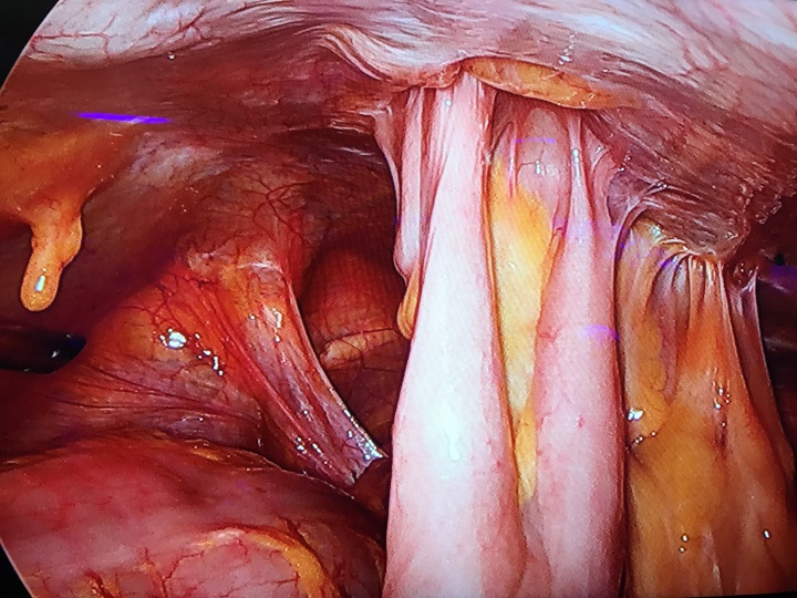
In the previous surgeries the risk of adhesions at umbilicus (Figure 2) could vary from 0% to 68% in those without any previous abdominal surgery, 0% to 15% in those with previous laparoscopy, 20% to 30% in those with previous laparotomy through a low transverse incision and 30% to 50% in those with a previous midline laparotomy.20–23 According to Ellis et al and Liakakos et al postsurgical adhesions are seen in 67–93% patients after general surgical operations and 70–95% patients undergoing major gynaecologic surgery.24,25

Figure 2 5mm port at Jain point and left side free of adhesions, whereas bowel loops adherent at the umbilicus.
Large masses which rise above the umbilicus, carry the risk of being directly hit by first blind trocar from umbilicus. In myomas it can cause bleeding and in case of cystic masses it can cause rupture of the cyst even before gaining access,26,27 and if malignant also carry the risk of upstaging the tumour due to spillage of cyst contents. Hence it will be prudent to use a non - umbilical approach. All this discussion clearly points to adopting a non-umbilical approach in more advanced situations. As we consider the concerns of major vessel injury via direct hit through veress or trocar or visceral injuries in case of bowel adhesions. May be, if we adopt the first blind entry through non-umbilical approach especially in vulnerable cases, we can avoid lot of complications due to the umbilical entry.
So, the authors thought goes with a newer concept of a non umbilical entry preferably lateral location. It lies in a large nascent area without any risk to major vessel or viscera and least chances of adhesions. So, by making a 5 mm trocar entry routinely through non – umbilical port and inspecting the abdomen 360° from liver, diaphragm right up to pelvis and then optimize the entry of 10 mm telescope port and the working ports according to mandate of the case and surgeon preference. Hereby we suggest newer lateral paraumbilical point – “Jain Point” described first in 2016.28 The surface marking defines it being on a vertical line drawn 2.5cm medial to Anterior Superior Iliac Spine, at the level of umbilicus. It has the advantage of having a fixed, very prominent bony landmark the ASIS (anterior superior iliac spine) making accurate entry possible. The veress needle and trocar are inserted in a straight vertical line, and there is no need to lift the abdominal wall making an easy simplified entry. This point is used first to introduce the veress needle and then after Pneumoperitoneum is created, 5mm trocar is inserted and whole abdomen is inspected 360° to assess adhesions and all other ports placed under visual control.
Author brought up this novel technique after their own experience for a decade on 7500 patients and publishing their experiences in various peer reviewed journals. Few other authors who adopted the same technique also suggest the need of non umbilical technique. Sharp et al also emphasized the need of non umbilical entry in low BMI patients and previous surgery cases.29 All these concerns prompted us to analyze an alternate entry method as a first blind non umbilical entry that we call “The Jain point”. Baris Mulayam et al.30 used this point in a patient with previous two caesarean sections with a Pfannenstiel incision, one laparotomic cholecystectomy with a Kocher incision, and a lumber disk operation. He used Jain Point for a direct trocar entry and concluded that there were no adhesion under Jain point (left lateral port) though adhesions were seen under the cholecystectomy scar till the umbilicus. There were no entry-related, intraoperative, or postoperative complications on entry through this point. Mohapatra and Bhusan31 have also reported the benefit of the this point as the main working port as well as the entry port, indicating dual benefit with good ergonomics. H.T.Sharp29 in upToDate proposed Jain point as an alternative non-umbilical site as it is “lower and more lateral in position compared with Palmer's point and may, therefore, be more easily used as the veress needle entry and then main operating port throughout the surgery.”He also considered it as a good operating port throughout the surgery.
The benefits of non umbilical ports are (Figure 3) moving away from adhesions at umbilicus, avoiding the major retroperitoneal vessels and safe entry in larger masses. So, the idea here is to introduce the concept on non–umbilical entry, avoiding umbilicus as RCOG32 also opines, umbilicus may not be the safest point of entry. The reader can formulate a plan to use either of the above described non – umbilical ports according to the clinical situation posed.
As the conventional non–umbilical techniques Palmer’s Point33 is most widely practiced for decades. The other two closely behind are Lee Huang Point34 and the 9th Intercostal Space.35 But none of these is either free of contraindications or complications. Hereby we suggest newer lateral paraumbilical point – “Jain Point” as per our practice and experiences.28,36,37
Lee Huang could be good for gynae malignancies and larger masses, being higher up than umbilicus. Palmer’s point turned out safe in many cases with dense abdominal adhesions but still contraindicated in upper abdominal masses and previous surgery scars, Splenic enlargement as in portal hypertension and bloated stomach.38,39 In previous mesh hernia repair, where the mesh is large and coming above the umbilicus, Palmer’s point may not be advisable as being more medial it can still come in the way of mesh. Jain Point may be considered in the situations of limitations of the conventional points especially Palmer’s Point, owing to its anatomical benefits being more lateral and lower down. There is comfortable vertical entry, so Jain point entry is easy to learn and continues to function as a main working port in due course of surgery. It is of value in previous surgery cases, large masses, extremes of BMI, the very thin and obese patients alike.
It is an overview to the concept of non-umbilical first blind port entry in the endeavour of reducing the major and minor complications which could be associated with first blind umbilical entry. Non-umbilical first blind entry of veress needle as well as of the first 5mm trocar entry through a non-umbilical approach holds promise to make laparoscopic entry safer. So, here the readers are given a different approach to laparoscopic entry in the pursuit of making laparoscopy safer. In using jain point as a routine practice of laparoscopic entry one can avoid injury to vessel, viscera, adhesions and bowel. This approach can also be used in limitations of conventional ports, like the palmers point.
None.
None.
All authors declare that they have no competing interests.

© . This is an open access article distributed under the terms of the, which permits unrestricted use, distribution, and build upon your work non-commercially.