eISSN: 2377-4304


Case Report Volume 16 Issue 5
 Khlifi Aymen,1 Aidi Hadhami,1 Dinari Amrou,1 Fdhila Sana,2 Dhouibi Maryam,1 Soui Slah3
1Gyneco-Obstetrics Department Kairouan, Tunisia
2Monastir Faculty of Medicine, Tunisia
3Department of Anesthesia and Intensive Care Kairouan, Tunisia
Correspondence: Marwen Nadia, Department of Obstetrics and Gynecology, Kairouan, Faculty of Medicine of Sousse, Tunisia
Received: October 11, 2024 | Published: November 6, 2025
Citation: Nadia M, Aymen K, Hadhami A, et al. Late diagnosis of ectopic pregnancy on a scar: what approach should be taken?. Obstet Gynecol Int J. 2025;16(5):170-172. DOI: 10.15406/ogij.2025.16.00807
Pregnancy on a cesarean scar is a rare form of ectopic pregnancy that can endanger the life and health of the patient, particularly due to the risks of hemorrhage or early uterine rupture. The clinical presentation is characterized by pelvic pain and/or variable intensity metrorrhagia in patients with a history of hysterotomy. The diagnosis is established through a transvaginal ultrasound. Different treatment options have overlapped, sometimes potentially to exacerbate the situation.
We discuss A 40-year-old pregnant woman with prior history of two cesarean sections found to have cesarean scar pregnancy at approximately 10 weeks’ gestation and underwent a laparotomy. The purpose of our study was to focus on the diagnostic difficulties, the therapeutic and evolutionary modalities of this disease.
Keywords: hemorrhage, hysterotomy, woman, cesarean scar pregnancy
An ectopic pregnancy lodged in the myometrium of a prior cesarean scar is known as a cesarean scar pregnancy (CSP).1 The incidence of cesarean scar pregnancies is estimated to be between 1 in 1800 and 1 in 2216. The literature contains only limited series and case reports.2 It is uncommon to suspect that a pregnancy on a cesarean scar exists, which leads to a diagnosis that may be more accurately described as an ongoing abortion or a cervical pregnancy. As a result, it may take longer to diagnose a cesarean scar pregnancy early on, which could lead to disastrous consequences. A case report of a patient who was admitted to treat a cesarean scar pregnancy is presented here. We will then discuss, in light of recent literature, the diagnostic and therapeutic methods available for better management of cesarean scar pregnancies.
This is a case report of a 40-year-old gravida 3, para 2, aborta 0 who initially presented with complaints of metrorrhagia and a positive beta-human chorionic gonadotropin (beta-hCG). In further discussion with the patient, she reported a history of crampy abdominal pain with 10 weeks of amenorrhea. Her obstetric history is significant for two cesarean sections ; the indications were breech presentation and respiratory distress due to a COVID infection. She had no additional past surgical history, and a first trimester ultrasound was not performed.
On presentation, the physical exam was notable for a soft, non-tender gravid abdomen with a well-healed Pfannenstiel scar. Vital signs were within normal limits, with a heart rate of 88 bpm and a blood pressure of 126/74 mm Hg. Ultrasound demonstrated a gestational sac with an embryo measuring 48 mm, corresponding to 11 weeks gestational age, that was implanted in the lower uterine segment, likely within the cesarean section scar. There was no myometrium seen surrounding the sac, no gestational sac in the cavity of the uterus or cervical canal, and a closed cervical canal (Figure 1). A magnetic resonance imaging (MRI) scan was refused by the patient for financial reasons. She was admitted to the hospital with a diagnosis of cesarean scar pregnancy for inpatient observation to determine optimal management. We decided to perform an open laparotomy due to the possibility of massive bleeding after obtaining the patient’s consent. The gestational sac was removed, as well as most of the trophoblastic tissue that was adherent and invading the wall of the lower uterine segment (Figure 2) (Figure 3). She was discharged in good condition 5 days after the operation.
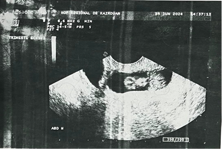
Figure 1 Endovaginal ultrasound photography showing the cesarean pregnancy scar.
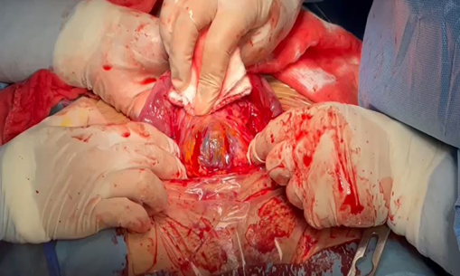
Figure 2 The bluish gestational sac was visible in the operating view, protruding from the dehiscent scar.
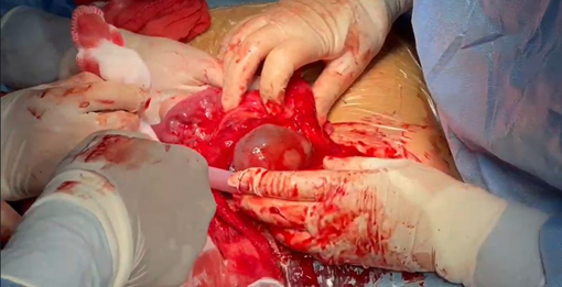
Figure 3 The bluish gestational sac was visible in the operating view, protruding from the dehiscent scar following a gentle incision of the fibro-muscular layer.
Scar pregnancy (SP) is a rare form of ectopic pregnancy, with incidence estimates ranging from 1 in 1800 to 1 in 2216 of all pregnancies.1 The first case was reported in 1978, and since then the number of reported cases has risen steadily, with an increased prevalence over the last two decades, secondary to an increase in primary and repeat cesarean sections. Additionally, during the COVID pandemic, there has been an increase in cesarean deliveries, with one study noting that 93% of COVID-positive pregnant patients underwent a cesarean section, and 61% of these had the procedure performed due to concerns about the effects of COVID-19 on the pregnancy.2
Its exact pathogenesis is unknown, but contributing factors have been thought to include endometrial and myometrial disruption or defects, as well as microscopic isolation between the cesarean scar and the space of the endometrium, leading to the implantation of the conceptus in the myometrium through the tract by the invading blastocyst.3 As the name suggests, a history of cesarean section remains the main predisposing factor, as in our case ; however, at present, there are no studies formally incriminating specific situations that expose the occurrence of this phenomenon.4
The clinical presentation is highly variable and can be misleading. In 39% of cases, there is metrorrhagia of varying severity (illustrated by our patient), isolated in a woman at the start of pregnancy with at least one previous cesarean section, which may be accompanied by pelvic pain in 16% of cases. This may be the main symptom in 9% of cases, and in a fairly representative 37% of cases, women may be asymptomatic; whereas the most dramatic situation is the combination of hemodynamic instability following an inaugural uterine rupture or an error in diagnosis and management.5
The published literature shows that up to 13.6% of cesarean scar pregnancies (CSPs) are wrongly diagnosed and managed as inevitable miscarriages with a low sac or cervical pregnancy1 leading to massive bleeding that can be life-threatening for the patient. However, mastery of ultrasound (endovaginal) semiology, which is the examination of first intention, can help thwart and limit the number of unrecognized cases. It has recently been proposed that Caesarean scar pregnancies should be differentiated into type 1 or "on the scar" and type 2 or "in the niche"6:
Other imaging examinations (three-dimensional ultrasound, magnetic resonance imaging (MRI)) will only be requested if there is diagnostic doubt during the two-dimensional ultrasound.
The treatment of CSP should be individualized on the basis of the clinical presentation, gestational age, type of pregnancy on imaging, and fertility preservation. Medical treatment using MTX is usually attempted systemically or locally before surgical treatment.7
Previous studies have reported that an earlier gestational age and faster diagnosis result in a higher success rate of conservative treatments with MTX. Local MTX therapy can increase the concentration of MTX in one area and reduce side effects compared to systemic therapy.5
Surgical treatment is not recommended except in cases where the conservative treatment fails due to high morbidity and poor prognosis or in cases of uterine rupture due to late diagnosis.8 Dilation and curettage, vacuum extraction method, hysteroscopy, laparoscopy, and laparotomy can be operative treatment options. Recent reports have suggested that hysteroscopy offers little benefit when compared with curettage alone. Laparoscopic procedures should be performed by a highly skilled surgeon, and vasopressin injection or laparoscopic coagulation or ligation of the uterine artery may be used to reduce bleeding.5 Interventional radiological techniques such as uterine artery embolization have been successfully used.
In our case, the diagnosis of CSP was delayed and thus the risk of bleeding was higher. There was no established definition of advanced or delayed diagnosed CSP .We opted for laparotomy (Figure 2) (Figure 3) for lack of technical facilities and patient’s refusal of methotrexate treatment.
The risk of recurrence of scar pregnancy remains unknown to this day, with articles putting it at between 4% and 15.6%, and in a study by Greshukhina, the recurrence rate was 40%.9 However, the consensus is that any planned pregnancy after a scar pregnancy should be scheduled with a safety interval of between 3 months and 1 or 2 years. It is recommended that an early ultrasound be performed to ensure that the scar is properly located while looking for any signs of placenta accreta throughout the pregnancy. Delivery should also be scheduled, and most authors agree that an iterative cesarean section should be performed in this patient.
Although the diagnosis of cesarean scar pregnancy (CSP) is rarely delayed, its treatment is important because it can be life-threatening. When the diagnosis is made earlier, the prognosis is good, and the risk of hysterectomy is relatively low. However, no standardized treatment for CSP has been established, and few cases have been reported. More cases and multicenter cohort studies are needed.
The authors are sincerely grateful to the patient for giving the permission to share this informative report.
Data availability: No data is associated with this article.
The authors received no specific funding for this work.
The authors declare no conflicts of interest.

©2025 Nadia, et al. This is an open access article distributed under the terms of the, which permits unrestricted use, distribution, and build upon your work non-commercially.