eISSN: 2377-4304


Case Report Volume 1 Issue 1
1Gynaecology, Leicester Royal Infirmary, UK
2Pathology, Leicester Royal Infirmary, UK
Correspondence: Suzanna Dunkerton, Leicester General Hospital, Leicester, Leics, UK, Tel 7979591333
Received: August 15, 2014 | Published: August 20, 2014
Citation: Dunkerton S, Pankhania NK, Johnson C, et al. Combined mucinous cystadenoma and carcinoid tumour of the appendix with coexistent features of an endometrioma: a case report. Obstet Gynecol Int J. 2014;1(1):5-7. DOI: 10.15406/ogij.2014.01.00002
Introduction: Endometriosis is defined as ‘the presence of endometrial glands and stroma outside the uterine cavity’. Appendiceal cancers are rare tumours of the gastrointestinal tract. There are some reported cases of these two disease processes occurring simultaneously within separate lesions. However, there are no reported cases of the two diseases occurring within the same entity. We report the unique case of an appendiceal cancer (combined mucinous cyst adenoma and carcinoid tumour) with coexistant histological features of an endometrioma.
Case report: A 36 year old nulliparous woman was referred to clinic with primary infertility, with an unremarkable past medical history. After routine infertility investigations, diagnostic laparoscopy was carried out and endometriosis was diagnosed. A suspicious lesion was also seen on the appendix. A right hemicolectomy and appendectomy was performed. Histological results showed mucinous cystadenoma with well differentiated carcinoid tumour and coexistent features of an endometrioma.
Discussion: An association between endometriosis and cancer has been well documented in literature. Existence with appendiceal cancers is rare. Carcinoids and cyst adenomas are both common types of appendiceal cancers, found incidentally or mimicking acute appendicitis. Occurrence in the same lesion is rare and unique to be found with features of an endometrioma.
Conclusion: This case illustrates the broad spectrum of appendiceal and endometrial disease. We hope to highlight the interesting asymptomatic presentation of this patient and therefore the importance of requesting routine histopathological analysis after appendicectomy.
Keywords: endometrioma, appendix, mucinouscystadenoma
Endometriosis is defined as the presence of endometrial glands and stroma outside the uterine cavity1,2 and is thought to affect up to 10% of women.3 Endometriotic lesions can be found anywhere in the body, common sites are the ovaries, pelvic peritoneum and fallopian tubes. It is less commonly found at the cervix, bladder, lungs and bowel.4
An association between endometriosis and malignancy has been well documented in literature despite reported controversy of the relationship.5,6 Brinton et al.7 evaluated a large cohort of Swedish women (20,868) with a diagnosis of endometriosis, were an increased risk of cancer was found; in particular ovarian, breast and haematopoietic malignancies. Reports of endometriosis associated with cancers of the bowel are rare.
Appendicular tumours are rare and account for only 0.4% of all gastrointestinal tumours.8 They are usually found incidentally or during investigations for other disease processes (as in our case), accounting for 1% of appendectomies.9
Reports of endometriosis associated with bowel cancer are rare. One similar case has been reported by Azordgean et al.,10 which documented the coexistence of a carcinoid tumour of the appendix and ileal endometriosis, each found at separate locations.10 This case presented with right lower abdominal pain mimicking acute appendicitis and was treated surgically with excision of the ileum and appendix. We describe the unique, not yet reported case of a 36 year old woman presenting with primary infertility, who was found to have a mucinous cyst-adenoma and carcinoid tumour of the appendix with co-existent endometriosis.
A 36 year old asymptomatic woman was referred with a 3 year history of primary infertility. Her periods occurred every 35-38 days with an average bleed and she engaged in regular unprotected coitus. No other personal or family medical history was noted and she led a healthy lifestyle as a non-smoker with minimal alcohol intake.
Routine infertility tests for herself and her partner were mostly normal. Except her ultrasound scan revealed 3-4 endometriotic cysts in the right ovary, the largest being 3.5cm in diameter with a small cyst also found in the left ovary. Endocervical swabs revealed Chlamydia which was promptly treated. It was decided to proceed with a diagnostic laparoscopy with tubal patency dye test and excision of endometriosis if appropriate.
Diagnostic laparoscopy found extensive uterovesical, peritoneal and ovarian endometriosis.
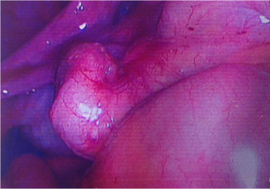
Figure 1: Right lateral bowel adhesions were dissected and endometriomas were excised from both ovaries and were mobilised as per RCOG guidelines.2 The dye test revealed the left tube was patent and the right blocked. Overall, grade 4 endometriosis was noted. On further visualisation of the abdomen, a suspicious looking mucoid lesion on the tip of the appendix was seen and biopsied.
Figure 2: Histology of the appendix tip showed mucinous material within the bowel wall and stroma suggesting a mucocele or mucinous tumour of the appendix. The patient was referred to lower gastrointestinal surgeons. A CT scan found it difficult to visualise the appendix but adjacent to the caecum a 10mm low attenuation lesion with mural calcification was noted suggestive of an appendicular mucocele. No pelvic or retroperitoneal lymphadenopathy was noted and no lesions were noted within the chest.

Figure 3: The patient underwent a right hemicolectomy during which further endometriotic deposits were seen. The appendix appeared abnormal as it was thickened and adherent to caecum with mucinous material extruding from the tip. Three proximal puckered lesions were noted in last 20cm of the terminal ileum but no other peritoneal disease was noted.

Histological results found the same appendix showed.
Figure 4 & Figure 5: Currently, the patient is well and conceived naturally whilst waiting to start IVF treatment.
Endometriosis is one of the most common diseases seen by gynaecologists. The prevalence is difficult to estimate as women can be asymptomatic or present with varying degrees of pelvic pain.11 Endometriosis is associated with infertility and has been identified in 38.5% of infertile women as opposed to 5.2% of fertile women.12
Appendicular tumours have diverse histology. Carcinoid neoplasms are the most common, accounting for 66% of all tumours of the appendix. They are comprised of enterochromaffin cells and contain many neurosecretory granules that release serotonin, histamine and prostaglandins.13 As with endometriosis, the incidence and prevalence is unknown, as the disease is mainly asymptomatic. The average time for a carcinoid tumour to become symptomatic is 9 years. Carcinoid tumours may present late and be associated with metastasis if found within the tip of the appendix.
The most common benign appendix tumours are mucinous cystadenomas. These tumours are composed of intestinal epithelium are dysplastic and secrete mucin, giving rise to a mucocele. This often causes the appendix to dilate and can present as acute appendicitis. The presence of a mucinous cystadenoma with associated mucin within the appendix wall increases the risk of psuedomyxoma peritonei.
There have been reports in literature of the co-existence of two types of appendicular tumours. Alsaad et al.14 reported a case of combined goblet cell carcinoid and mucinous cyst-adenoma of the appendix in a patient presenting with right iliac fossa pain. The occurrence of appendicular tumours co-existing with endometriosis is rarely reported,15 and there have been no reported cases of two different appendicular tumours being found with co-existent features of an endometrioma within the same appendix. Appendicectomy specimens should be routinely sent for histopathological analysis. Duzgan et al.15 reported an intra-operative detection rate of less than 50% for all types of appendicular tumours. Jones et al (2007) evaluated the histopathological reports of 1225 appendicectomy specimens. Of the 1225, 46 (3.75%) revealed abnormal diagnoses and 24 (1.96%) were clinically significant, altering patient management.16 Routine histopathological analysis avoids the potential to miss significant pathologies which may need different treatments.
Currently in the literature there are no clear guidelines regarding the treatment of appendicular tumours.17 There is considerable debate as to the use of appendectomy versus right hemicolectomy and the effects on long term results. A right hemicolectomy is indicated with adenocarcinomas, tumours invading the mesoappendix, serosa, lymphatics or vasculature and benign tumours with a diameter of more than 2cm.18 As preferred by some surgical units19 a right hemicolectomy is considered the treatment of choice. There was suspicion of a malignant adenocarcinoma after initial biopsy which rendered a right hemicolectomy a safer option.
Most surgeons will only encounter a few carcinoid tumours of the appendix in their career. We feel this unique case will add to the small database of similar reports and help with management of this rare disease.
This case illustrates the broad spectrum of appendicular and endometriotic disease. It also demonstrates the role of biopsy of abnormal areas atypical of endometriosis at diagnostic laparoscopy. We hope to highlight the interesting asymptomatic presentation of this patient and therefore the importance of requesting routine histopathological analysis. After prompt treatment with right hemicolectomy, this young woman has fortunately conceived naturally without IVF.
None.
Author has no any conflict of interest to declare.

©2014 Dunkerton, et al. This is an open access article distributed under the terms of the, which permits unrestricted use, distribution, and build upon your work non-commercially.