eISSN: 2377-4304


Case Report Volume 5 Issue 2
1Department of Obstetrics and Gynaecology, KK Women's and Children's Hospital, Singapore
2Department of Pathology, Singapore General Hospital, Singapore
3Department of Gynae-oncology, Singapore General Hospital, Singapore
Correspondence: Janice Su Zhen Tung, Department of Obstetrics and Gynaecology, KK Women's and Children's Hospital, 100 Bukit Timah Road, Singapore 229899
Received: September 04, 2016 | Published: September 26, 2016
Citation: Tung JSZ, Lih WS, Kheng CG. Case report of vulvar pyoderma gangrenosum in a BRCA 2 carrier. Obstet Gynecol Int J. 2016;5(2):310-312. DOI: 10.15406/ogij.2016.05.00153
We report a case of vulvar pyoderma gangrenosum which we successfully recognized and treated in a young female BRCA 2 carrier. Our patient’s presentation appears to be classical of pyoderma gangrenosum, specifically the occurrence of pathergy, and that of recurrent small boils rapidly evolving into angry ulcers. Histological hallmarks in the biopsy include a predominantly neutrophilic infiltrate and prominent vasculitic elements. Literature review finds that this is a rare condition that may be associated with underlying solid malignancies such as breast cancer. The BRCA 2 association in our patient may suggest a possible, yet obscure, genetic and immune pathogenesis link between pyoderma gangrenosum and certain cancers. On the immune basis, first-line treatment with steroids is usually effective. Vulvar pyoderma gangrenosum is an important but difficult diagnosis. It should be considered in recurrent or refractory genital ulcers, which can be debilitating in a young woman. Multidisciplinary team input is highly recommended.
Keywords:Genital ulcer, Vulvar pyoderma gangrenosum, Genetics, Immuno-pathogenesis, BRCA
We report a case of vulvar pyoderma gangrenosum diagnosed in a young female BRCA 2 carrier, after she presented with recurrent vulvar ulcerations. Literature review finds that this is a very rare, but important genital condition with an immuno-pathogenesis that maybe associated with underlying malignancies. It should be considered in a patient with recurrent or refractory genital ulcers.
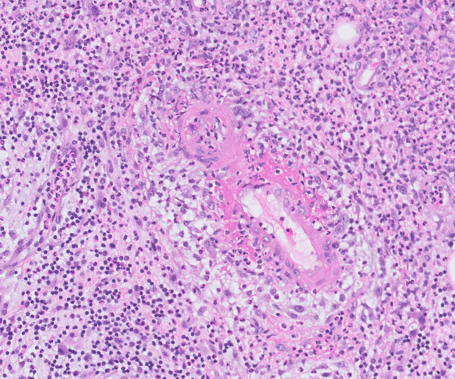
A 30-year-old single woman with no significant medical history first presented with a large necrotic right vulvar ulcer of 4 days’ duration after a history of saddle trauma. She underwent saucerisation with gradual healing of the wound over 1 month. Histology returned as an acute epidermal ulceration extending to the subcutaneous layer (Figure 1), with florid inflammatory, predominantly neutrophilic, perivascular infiltrate and myointimal proliferation, suggestive of a vasculitic element (Figure 2). No malignancy was seen. She was referred for follow-up with a specialised vulva clinic. In the subsequent year, similar ulcers recurred at the same site three to four months apart. A swab culture from the first recurrence was positive for Staphylococcus aureus. She was treated with antibiotics and wound care. The recurrent ulcers were slow to heal. Her blood count was normal. Evaluation for diabetes and venereal infections including chlamydia and herpesviruses negative. With each episode, the patient gave a history of sudden onset of a small boil that would grow rapidly and erupt into a sloughing painful blister with raised lilaceous edges. They tend to be triggered by particularly stressful periods in her life. There was no constitutional symptoms or involvement of other systems. She became depressed, tentative about physical examinations and declined further testing.

Figure 2 Predominantly neutrophilic leukocytoclastic vasculitis with fibrinoid necrosis at the edge of the lesion.
In her fourth recurrence, a multidisciplinary team involving rheumatology, dermatology and gynae-oncology reviewed her. Autoimmune differentials including Behcet’s disease and inflammatory bowel disease (Crohn’s and ulcerative colitis) were contemplated. Vulvar pyoderma gangrenosum was eventually diagnosed given her classical clinical presentation, the histopathological findings as described, and the negative workup for other differentials. She made a rapid recovery after commencing on a reducing regime of oral prednisolone over 2 months, and then subsequently, maintained on topical clobetasol for another 4 months. Her condition had since remained quiescent for more than a year after stopping treatment. This patient had a significant family history of breast and ovarian cancer. Her mother had both breast and ovarian cancer at the age of 45, and her maternal aunt had ovarian cancer. She also had a small right breast lump. Biopsy confirmed benign fibrocystic change. She agreed to a genetic screening and was found to be a BRCA 2 carrier, for which she received counselling accordingly.
The patient chose expectant management with regards to her increased risks for breast and ovarian cancer, and declined risk-reducing surgery. A discussion into her acceptance for physical intimacy and reproductive health outlook revealed that she was not considering starting a family. She had concerns about knowingly transmitting the mutation to her daughters and subjecting them to the same risk management dilemmas of a carrier. Currently, preimplantation genetic diagnosis involving BRCA mutation carriers is not performed in Singapore.
Recurrent genital ulceration represents a diagnostic challenge. Differentials include infectious, autoimmune or malignant processes. Venereal infections such as herpes may give rise to painful genital ulceration, however, they are unlikely to appear as large purulent necrotic ulcers with over-hanging borders. Secondary infections may occur for which antibiotics may appear to effect a response. Other autoimmune processes such as metastatic Crohn’s disease and Behcet’s disease need to be considered. These diagnoses were unlikely in this patient however, as she had isolated genital lesions, without bowel symptoms, oral aphthosis, uveitis and other skin or vascular manifestations (e.g. phlebitis, thrombosis). Malignancy mustal ways be excluded in slow-healing or recurrent ulcers. Our patient’s presentation appears to be classical of pyoderma gangrenosum, specifically its occurrence at the same site as previous trauma1 exhibiting the phenomenon of pathergy, and that it would begin as a small boil that would rapidly evolve into an angrier ulcerative necrotic picture. Clinical presentations of the condition described in literature2 are usually likened to its term “pyoderma gangrenosum”, although the actual pathology is neither infectious nor gangrenous.
Currently, no established diagnostic criteria or pathognomonic histopathological features for vulvar pyoderma gangrenosum exist. However, the literature has suggested a prominent vascular component, such as that in our biopsy, as a possible hallmark in its histology2 The histological pictures (Figure 1 & 2) demonstrate the dense neutrophilic infiltrate and perivascular cuffing of leukocytoclastic vasculitis, with fibrinoid necrosis, at the edge of the lesion. In contrast, benign aphthous ulcers usually contain a mononuclear type of infiltrate with a fibrin coating,3 although their pathophysiology remains as uncertain as that of pyoderma gangrenosum ulcers.
Aetiology of pyoderma gangrenosumis poorly understood, with immune dysregulation theories simplicated. It seems to manifest as a paraneoplastic dermatosis4 in some cases and has been associated in 50% to 70% of cases with solid malignancies including breast cancer5 and other hematologic malignancies.6 In the absence of malignancy, extracutaneous involvement of pyoderma gangrenosum has also been known to occur and can be fatal, as with a case report of vulvar pyoderma gangrenosum with pulmonary infiltrates.7
Anatomically, since primitive embryonic milk lines extend from axilla to groin, the vulva has been popularly associated with mammary cancers,8 whether that of primary synchronous breast and vulvar cancers, metastatic breast cancer to the vulva, vulvar cancer metastatic to the breast,9 cancer of ectopic supernumerary breast tissue in the vulva10 or extra mammary Paget’s disease.
The BRCA 2 mutation detected in this patient is of special interest. A recent molecular report demonstrated that BRCA 2 deficiency in mouse models could play a role in immune dysfunction.11 A possible genetic and immune link in the pathogenesis of pyoderma gangrenosum and its association with certain cancers may exist. It is yet obscure and remains to be addressed with further studies. Cases of pyoderma gangrenosum occurring at the breast wound site after surgical treatment of breast cancer and breast reconstruction surgery12 have been reported in the literature. One of them is the only other published case involving a BRCA carrier (BRCA 1).13 It is uncertain if the development of this cutaneous manifestation will significantly modify the risks inherent in this patient’s carrier diagnosis.
On the immune basis, first-line treatment with systemic steroids is usually effective. Other immunosuppressive agents including cyclosporine or azathioprine may be employed as steroid-sparing agents in severe disease. Targeted therapy with immuno-modulating monoclonal antibodies such as infliximab is, at present, experimental. There has been an association between use of Rituximab in the treatment of lymphoma and the development of vulvar pyoderma gangrenosum itself.14 Aggressive surgical debridement may result in pathergy and wound enlargement, and should generally be avoided. Meticulous wound care as the ulcer heals is paramount. We can consider suppression of menstruation to facilitate perineal hygiene while lesions are active, after weighing the risks of short term hormonal treatment in a BRCA 2 carrier. Topical super potent steroids may help in limiting recurrence after recovery.
As with other vulvar diseases, this disfiguring and painful condition can be debilitating for a young woman. We must learn to recognize it early in order to institute prompt treatment, avoid complications and minimize its psychological and social impact on patients. Specialist follow-up and multidisciplinary team involvement are recommended. We should remain sensitive and perceptive towards the woman’s feelings and concerns with regards to her sexual and reproductive health.
We would like to acknowledge Dr. Rafay Azhar’s contribution in providing the histopathology slides for this article.
None.

©2016 Tung, et al. This is an open access article distributed under the terms of the, which permits unrestricted use, distribution, and build upon your work non-commercially.