eISSN: 2377-4304


Case Report Volume 13 Issue 5
1Obstetrics and Gynecology, ABC Medical Center, Instituto de Estudios Superiores de Monterrey, Mexico
2Obstetrics and Gynecology, ABC Medical Center, Universidad Nacional Autónoma de México, Mexico
3Obstetrics and Gynecology, ABC Medical Center, Universidad Nacional Autónoma de México/Universidad Anáhuac, Mexico
Correspondence: Ayala-Yáñez Rodrigo, ABC Medical Center, Mexico City, Mexico
Received: August 31, 2022 | Published: September 23, 2022
Citation: DOI: 10.15406/ogij.2022.13.00666
Breast cancer in male patients is a rare clinical entity, representing less than 1% of all cancer incidence in males. Breast cancer is highly associated to hyperestrogenism, obesity and alcohol intake, and it is a disease mostly considered in female patients, hence, the lack of suspicion and proper evaluation in male patients. It must be considered that these tumors may be malignant and require an adequate protocol to avoid establishing diagnosis in advanced stages. This is the case of a 19-year-old male in Mexico City, who was diagnosed with a fibromixoid sarcoma, based on mastography and ultrasound imaging, biopsy and immunohistochemical signature. Tumor was excised through radical left breast mastectomy with final histologic diagnosis being fibromixoid sarcoma (T2a N0 M0), clinical stage IB. No further treatment was required, and periodical outpatient consultations are performed for follow up. The most significant risk factor found in our patient was an increased body mass index (BMI), smoking and alcohol consumption.
Keywords: fibromixoid, sarcoma, male, breast, cancer
Breast cancer is a global health problem, great efforts and resources are destinated to make an early diagnosis and proper treatment, still, this pathology is rare among male patients and therefore, widely understudied.1 Comparing the incidence in both genres, breast cancer represents less than 1% of all cancers affecting male patients, compared to 12.5% in females (CDC Reports a rate of 1.28 per 100,000 population vs. 125.11 in 100,000 population).2–4 Breast sarcomas are rare in both groups, it is less than 5% of the overall sarcoma cases and it represents only 1% of total breast cancer in female patients.5 Breast sarcomas are a heterogeneous group of non-epithelial tumors derived from mesenchymal tissue6,7 yet, actual etiology remains elusive and poorly understood in males. Presentation mean age in female patients is 62, while for males it ranges between 67 and 69 years of age,2 androgen positive receptor tissue is present in 97% of male breast cancer while females only report 61%, and BRCA mutations are infrequent in male patients. Other risk factors to consider in these patients is family history for breast cancer (OR: 3.65; 95% CI:1.62-8.19), and high body mass index of 31kg/m2 (BMI) (OR:1.6;95% CI: 0.85-3.02). Moderate physical activity seems to be a protective factor, still, this is based on information provided from small cohorts and requires further research.8 We present the case report of a young male in Mexico with breast cancer.
A 19-year-old male who in the last 4 months has had progressive left breast growth and a palpable mass, he did not report any swelling or bleeding. He had no prior medical or surgical conditions though he did have a history of tobacco smoking and moderate alcohol consumption. Family history was positive only to liver and gastric cancer. Physical examination revealed severe gynecomastia, a moveable mass in the left breast, approximately 2-3cm in diameter, located between 1:00 and 2:00 o’clock radius, line B, with significant skin color, and texture changes, no adenopathy was detected.
The mass was studied with mammography revealing fibroadipose tissue and a heterogenic nodule with amorphous calcifications and well-established margins in the upper outer quadrant. Mammary ultrasound confirmed the fibroadipose texture and the nodule with a heterogeneous content, dimensions were determined to be 30.3x27.4x22.8mm, the study was staged as Breast Imaging Reporting and Data System 4A (BIRADS). A Tru-Cut biopsy was performed, finding a stromal lesion with myofibroblast differentiation. As for the immunohistochemical signature, the lesion was negative for estrogen receptor (RES), negative diethylstilbestrol exposition factor (DES), positive for cluster differentiation 99 (CD-99) and negative for cluster differentiation-34 (CD-34). The lesion was diagnosed as a fibromixoid sarcoma of the left breast T2a N0 M0, clinical stage IB (FIGO Breast Cancer Classification 2019).
Left sided radical mastectomy was performed without any complications, findings were a solid mass of 6 x 4 x 3 cm, surgical margins were negative for malignant cells. Final immunohistochemical analysis reported positive cluster differentiation 68 (CD-68), positive B cell lymphoma 2 (BCL-2), negative S-100, and positive Ki67 (only in 5%). Final diagnosis being a fibromixoid sarcoma. No chemo or radiotherapy was required since it was a low-grade lesion. Outpatient follow-up has continued without further developments.
Our patient’s main risk factor was his increased BMI (>30 kg/m2), smoking and alcohol consumption and family cases of hepatic and gastric carcinoma, although these factors are associated to cancer risk, they are not specific for this specific sarcoma.9 Sarcomas are associated to molecular anomalies in BRCA and P53, among other genes.10 Due to the low incidence and low odds ratios encountered in these tumors, there is a possibility that environmental (radiation) and family hereditary factors may hint a multifactorial origin. Some articles mention the exposition to ionizing radiation, smoking and other factors as a possible risk (seen only in 0.2% of cases),7,10,11 though our patient had no prior radiation exposure.
Fibromixoid sarcomas are soft tissue tumors with fibroblast cells with a circular pattern and collagen deposits, immunohistopathologic markers include MUC 4 glycoprotein, EMA, S100, CD34, vimectin, desmin, and keratin.12,13 There are no characteristic radiological imaging features and clinical staging is based on the TNM system, American Joint Committee on Cancer System (AJCC).11,14 This patient had a low malignancy tumor since it was smaller than 5 cm and with scarce vascularity (both, if present, are considered as high malignancy potential).15 Current treatment is surgical with a total mastectomy, postoperative treatments like radio or chemotherapy are based on the presence of malignant cells in the tumor borders upon excision, although sarcomas do have a high recurrence rate. These tumors don’t have lymphatic dissemination; hence, no lymphatic resection is required (Figures 1–3).16,17
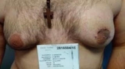
Figure 1 Patient’s breasts reveal a advanced gynecomastia condition, with normal android hair distribution in chest. Left breast reveals significant superficial changes around the areola with scarring and deformation. No pain or secretion was referred, still movable mass was easily found with manual exploration.
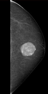
Figure 2 Left breast mammography reveals a round heterogeneous nodule, located between 1:00 and 2:00 o’clock radius, line B, dimensions were determined to be 30.3 x 27.4 x 22.8 mm, the study was staged as Breast Imaging Reporting and Data System 4A (BIRADS).
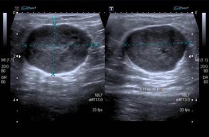
Figure 3 Left breast nodule ultrasonography, reveals a heterogeneous, avascular lesion located at 1:00 and 2:00 o’clock radius, line b.
Breast cancer in male patients is rare and may go undiagnosed due to lack of suspicion of a probable malignant tumor in these cases, much of their clinical behavior is unknown and based on few cases. All breast tumors, independent of gender should be properly examined and tested due to the probability of finding a malignant tumor involved.
None.
None.
None of the authors have any conflict of interest.

©2022 , et al. This is an open access article distributed under the terms of the, which permits unrestricted use, distribution, and build upon your work non-commercially.